Sticta fuscotomentosa B. Moncada, Coca & Lücking, in Ariyawansa et al., Fungal Diversity: 10.1007/s13225-015-0346-5, [111] (2015)
Index Fungorum number: IF551488; Facesoffungi number: FoF: 00929
Etymology – Referring to the superficial similarity with Sticta tomentosa.
Holotype – L.F. Coca 1207 (FAUC).
Diagnosis – Differing from Sticta tomentosa and S. leucoblepharis in the brown to dark tomentum and the 2–4 papillae on the cells of the basal membrane of the cyphellae.
Thallus epiphytic, corticolous. Primary photobiont a cyanobacterium(Nostoc). Basal stipe absent or indistinct. Thallus palmate to irregular, up to 10 cm across, moderately branched. Lobes laciniate to ligulate, ascendant to erect, adjacent to imbricate, plane to involute, with rounded tips; margins straight to sinuose; lobe internodes 4–12 mm long and (5–)10–20 mm broad. Upper surface even to shallowly foveolate (pointimpressed), dark green when fresh and bluish grey in the herbarium, opaque to slightly glossy, glabrous, with scattered, irregular, cream-coloured maculae; cilia abundant, dark greyish brown, up to 1 mm long. Vegetative propagules absent. Apothecia abundant, mostly laminal, substipitate, up to 1.3 mm diam. and 330μm high; disc reddish brown; margin
entire, cream-coloured. Excipulum up to 120μm broad; hymenium up to 100μm high; epithecium up to 5μm high, orange. Ascospores fusiform, 1–3-septate, 20–35×7–8μm, hyaline. Medulla loose in surface view, whitish to creamcoloured, K+ yellow, C–, KC–, P–. Upper cortex paraplectenchymatous, 20–30μm thick; upper layer with smaller, thicker-walled cells and lower layer with larger, thinner-walled cells; photobiont layer 10–20μm thick; medulla 75–120μm thick, with yellow-orange crystals; lower cortex paraplectenchymatous, 15–20μm thick. Lower surface even to slightly undulate, whitish to cream-coloured, but becoming darker towards the center. Primary tomentum irregular, thick, becoming sparse and thin towards the margin, arachnoid to spongy, beige to dark greyish brown towards the center, hairs 120–750μm long, forming fascicles of 12–20 unbranched, septate hyphae; secondary tomentum absent. Rhizines absent. Cyphellae abundant, 1–20 per cm2 towards the center and 41– 60 per cm2 towards the margin, dispersed, angular-rounded, urceolate with wide pore, (0.25–)0.4–0.7(−1.3) mm diam., immersed to prominent but remaining below the level of the tomentum, with involute, whitish to cream-coloured, glabrous margin; basal membrane finely pubescent, with 2–4 papillae per cell, pale yellowish, K+ yellow, C–, KC–, P–. Pycnidia not observed. Secondary chemistry: No substances detected by TLC but medulla and basal membrane of the cyphellae with dispersed, orange-yellow pigment granules in anatomical sections.
Material examined − COLOMBIA, Risaralda, Mun. Santuario, Tatamá National Natural Park, close to Monte Zancudo, 2600 m, 13 January 2011, L.F. Coca et al. 1207 (FAUC holotype; UDBC isotype); Quindío, Mun. Salento, Navarco Alto trail, Piscícola station, trail to the line, 3000 m, 4 November 1991, J. Uribe & A. Bonilla 2173 (COL); Risaralda, Mun. Pereira, Otún Quimbaya Fauna and Flora Sanctuary, La Florida, La Suiza trail, 1820 m, 2 September 2003, B. Moncada
& R. Dávila 1908b (UDBC); Tolima, Mun. Ibagué, Las Juntas, El Silencio to El Rancho road, 2600 m, 18 May 2008, B. Moncada 2531b (UDBC).
Distribution and Ecology − Sticta fuscotomentosa is thus far known from the Colombian Andes, in the upper montane cloud forest zone between 1820 and 3000 m altitude. The species is typically found epiphytically in shady to semiexposed microsites.
Notes − Sticta fuscotomentosa belongs in the S. tomentosa clade (Moncada et al. 2014a, b; Fig. 68) and is most similar to S. tomentosa (Sw.) Ach. and S. leucoblepharis (Nyl.) Tuck. & Mont. and differs chiefly in the dark lower tomentum towards the thallus center and the dark marginal cilia (Moncada 2012). The apothecial margin is glabrous, whereas young apothecia in S. tomentosa and apothecia in S. leucoblepharis have tomentose margins.
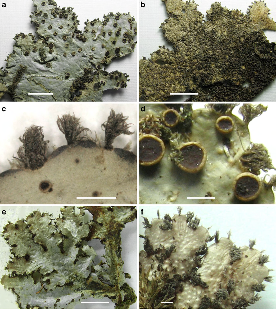
Fig. 1 Sticta fuscotomentosa (holotype) a Lobe upper side with apothecia b Lobe underside showing dark tomentum c Marginal cilia enlarged d Apothecia enlarged. Sticta subfilicinella (holotype) e Lobe upper side f Lobe underside showing marginal cilia and tomentum. Scale bars: a–b, e=10 mm, c–d, f=1 mm