Septomelanconiella thailandica Samarak. & K.D. Hyde, sp. nov.
Index Fungorum number: IF555302
Etymology: Name based on the country from which this species was collected, Thailand.
Holotype: MFLU 18-0793
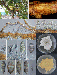
Saprobic on recently dead twigs of Syzygium samarangense. Sexual morph: Undetermined. Asexual morph: Coelomycetous. Conidiomata 360–500 μm diam., 130–200 μm high, pycnidial, immersed, partially erumpent at maturity, solitary or confluent, subglobose to irregular, to flattened and collabent, light brown. Conidiomata walls 28–37.5 lm wide, comprising 2–3 layers of hyaline cells, of textura angularis at the base, with light brown, thin outer layer. Conidiophores mostly reduced to conidiogenous cells, few with conidiophore and cylindrical conidiogenous cells. Conidiogenous cells 10.3–15. × 4.4–7.3 μm (x̅ = 13.2 × 5.9 μm, n = 20), enteroblastic, phialidic, integrated or discrete, cylindrical, determinate, hyaline, finely roughened. Conidia when immature, cylindrical to clavate, one guttulate, becoming two guttules, hyaline, one septate; mature conidia 37–52 × 15–23 μm (x̅ = 43.8 × 19.1 μm, n = 40), cylindrical to clavate, straight or slightly curved, brown, 1-euseptate, with 6 unequal lumina, guttulate, dark brown at base with opening 1.8–3.6 μm diam. (x̅ = 2.9 μm, n = 30).
Culture characteristics: Conidia germinating on PDA within 24 h, germ tubes produced from central part, often three hyphae. Colonies on PDA reaching 37 mm diam. in 2 weeks at 25 °C, hairy, white, superficial, rough surface, irregular edge, reverse yellowish brown.
Material examined: THAILAND, Chiang Rai Province, Muang District, Nang Lae, on dead twigs of Syzygium samarangense (Blume) Merr. & L.M. Perry (Myrtaceae), 25 January 2018, M.C. Samarakoon, SAMC091 (MFLU 18-0793, holotype; KUN-HKAS 102320, isotype), ex-type living culture, MFLUCC 18-0518, ICMP. GenBank numbers: ITS = MH727706, LSU = MH727705, RPB2 = MH752072.

Fig 1. Septomelanconiella thailandica (MFLU 18-0793, holotype). a Host. b Conidiomata on the substrate. c Vertical section of conidioma. d–h Conidiophores, conidiogenous cells and developing conidia. i–l Conidia. m Germinated conidium. n Culture on PDA from above after 16 days. o Culture on PDA from below after 16 days. Scale bars b = 1000 μm, c = 100 μm, m = 50 μm, d–l = 20 μm.
