Scolecoleotia eriocamporesi H.B. Jiang, Phookamsak & K.D. Hyde, sp. nov.
MycoBank number: MB 558193; Index Fungorum number: IF 558193; Facesofungi number: FoF 09764; Fig. 115
Etymology: Named after Erio Camporesi who has con- tributed many fungal collections from Italy.
Holotype: MFLU 16-2133
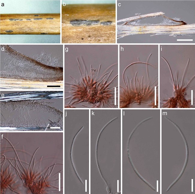
Saprobic on dead aerial fronds of Pteridium aquilinum. Sexual morph Undetermined. Asexual morph Coelomycetous, visible as black, raised, elongate area on the host. Conidiomata 75–145 μm high, 600–1100 μm long, black, pycnidial, solitary to gregarious, scattered, sometimes arranged in rows on host substrates, immersed, slightly raised, elongate, hemisphaerical to subconical, with wedge-shaped at the basal angles, uniloculate, glabrous, apapillate, with inconspicuous ostiolate. Pycnidial wall 10–30 μm wide, thin-wall of equally thickness, composed of 3–5 layers, of brown to dark brown pseudoparenchymatous cells, arranged in textura angularis to textura globulosa, difficult to distinguish from the hymenium. Hymenium composed of 2–3-strata, of hyaline polygonal cells, polygonal cells 3–6 × 3–6 μm (x̅ = 5 × 5 μm, n = 30). Conidiophores reduced to conidiogenous cells. Conidiogenous cells (5.5–)7–10(–12) × 2–3(–4) μm (x̅ = 9 × 3 μm, n = 50), holoblastic, phialidic, determinate, discrete, hyaline, ampulliform to subcylindrical, aseptate, smooth-walled, arising from the innermost layer of cavity of conidioma. Conidia (23–)40–65(–75) × 1.5–2.5 μm (x̅ = 57 × 2 μm, n = 50), acrogenous, scolecosporous, solitary, cylindrical to filiform, curved, hyaline, aseptate, smooth-walled.
Material examined: ITALY, Province of Arezzo, Montemezzano-Stia, on dead aerial fronds of Pteridium aquilinum, 7 July 2016, E. Camporesi, IT3027A (MFLU 16-2133, holo- type); ibid., MFLU 16-2133 (isotype).
GenBank numbers: ITS = MW981448, MW981449, LSU = MW981450, MW981451, SSU = MW981452, MW981453.
Notes: Based on the NCBI BLASTn search results of the ITS, LSU and SSU sequences, Scolecoleotia eriocamporesi (strains IT3027A and IT3027B) showed 92% similarity with Urceolella carestiana (CBS 319.71), 96.26% similarity with Leotiomycetes sp. (BY-2018b) and 99.36% similarity with Dicephalospora rufocornea (MFLU 18-1827), respectively.

Fig. 115 Scolecoleotia eriocamporesi (MFLU 16-2133, holotype). a, b Appearance of conidiomata on host surface. c Section through con- idioma. d, e Section through pycnidial wall. f–i Conidiogenous cells with attached conidia stained in Congo red. j–m Conidia. Scale bars: c = 500 μm, d, e = 50 μm, f–i = 20 μm, j–m = 10 μm
