Sarimanas shirakamiense Matsumura, K. Hiray. & Kaz. Tanaka.
Index Fungorum number: IF551054, Facesoffungi number: FoF00497; Fig. 2
Etymology – In reference to the location where the specimen was collected.
Holotype – HHUF 30454
Saprobic on Swida controversa. Sexual morph Ascomata 220 – 300 μm high, 200 – 320 μm diam., mostly scattered, sometimes 2 – 3 gregarious, immersed, globose in section. Neck 30 – 45 μm high, 70 – 80 μm wide, short papillate, without clypeus. Peridium uniformly 25 – 30 μm thick, composed of 4 – 6 layers of polygonal, 10 – 20 × 4 – 8 μm, thin-walled cells. Hamathecium numerous, cellular, 2 – 3 μm wide, hyaline, septate, branched and anastomosed. Asci 95 – 158 (–175) × 26.5 – 52.5 μm (x̄ = 133.2 × 39.6 μm, n = 30), 8 – spored, not numerous, bitunicate, fissitunicate, cylindrical to ovoid, with a short pedicel (8.5 – 20 μm) long, apically rounded with an ocular chamber. Ascospores (30–) 32 – 51 × 10 – 17 (–18.5) μm (x̄ = 40.3 × 13.9 μm, n = 60), L/W 2.2 – 3.8 (x̄ = 2.9, n = 60), broadly fusiform with rounded ends, mostly straight, thick-walled, with a septum mostly submedian (0.49 – 0.62; x̄ = 0.53, n = 60), hyaline, smooth-walled, guttulate when young, with an entire sheath; sheath when fresh condition diffuse, gelatinous, up to 8μm wide, later becoming sharply delimited firm sheath of 2 – 4 μm thick. Asexual morph Undetermined.
Culture characters – Colonies on PDA, white, buff pigment produced; on MEA, white to olivaceous grey; on CMA, white to lavender grey. On RSA, numerous ascomata are produced on the surface of rice straw. Ascomata ca. 400μm high, ca. 250μm diam., immersed, globose, with papilla. Asci and ascospores are considerably larger than those on the natural specimen; asci 162–225×34–46.5μm (x̄ = 192.8 × 38.5 μm,n = 20); ascospores (42.5–) 45 – 51 (–53) × (10–) 15 – 17.5 μm (x̄ = 47.5 × 15.4 μm, n = 50), L/W (2.6–) 2.8 – 3.3(–3.6) (x̄ = 3.1, n = 50), with a septum mostly submedian (0.48 – 0.54; x̄ = 0.51, n = 50).
Material examined – JAPAN, Aomori, Nishimeya, Shirakami Natural Science Park of Hirosaki Univ., on twigs of Swida controversa (Cornaceae), 3 June 2012, K. Tanaka, KT 3000 (HHUF 30454, holotype designated here); ex-type living culture, MAFF 244768. GenBank ITS: LC001718; LSU: LC001715; SSU: LC001712; ibid., Aomori, Nishimeya, Seisyu-trail, on dead twigs of woody plant, 29 May 2007, K. Hirayama et al., KH 13 (HHUF 30081, paratype); ex-paratype living cultures, JCM 17825, MAFF 242969. GenBank ITS: LC001719; LSU: LC001716; SSU: LC001713.
Notes – A new genus, Sarimanas, is established for S. shirakamiense and S. pseudofluviatile based on their shared morphological characters, such as globose to subglobose ascomata composed of polygonal thin-walled cells, cylindrical to ovoid asci with a short pedicel, and broadly fusiform, 1 – septate, hyaline ascospores with an entire sheath. These morphological features of Sarimanas are somewhat similar to those of Massarina, but the latter genus has clypeate ascomata and belongs to Massarinaceae (Hyde et al. 1999; Zhang et al. 2012). Ascospore morphology of Sarimanas superficially resembles to that of Wettsteinina in Pleomassariaceae (Kodsueb et al. 2006) or Lentitheciaceae (Schoch et al. 2009), but the latter genus has ascomata lacking a hamathecium when mature (Zhang et al. 2012a, b). The phylogenetic analysis based on LSU(Fig. 1) suggested that this genus has close affinity to genera in Melanommataceae, such as Aposphaeria and Herpotrichia, but within in this family no genus phenotypically similar to Sarimanas is known. Sarimanas shirakamiense is similar to S. pseudofluviatile, but the asci and ascospores of S. shirakamiense are considerably larger than those of the latter (asci 99 × 20.2 μm, ascospores 30.6 × 8.7 μm in average). Sequence differences between these two taxa were found at eight positions in the ITS region, suggesting that they are not conspecific.
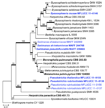
Fig. 1 Phylogram generated from Maximum likelihood (RAxML) analysis based on combined LSU, SSU and TEF1 sequence data of Melannomataceae. Maximum likelihood bootstrap support values greater than 50 % are indicated above or below the nodes, and branches with Bayesian posterior probabilities greater than 0.95 are given in bold. The ex-types (reference strains) are in bold; the new isolates are in blue. The tree is rooted with Biatriospora marina strain CY 1228.
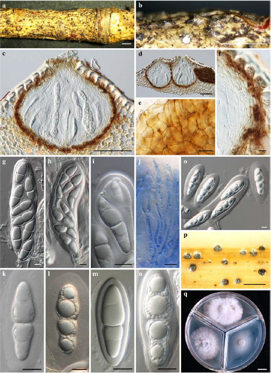
Fig. 2 Sarimanas shirakamiense (holotype). a, b Ascomata on host surface c, d Ascomata in longitudinal section e Peridium of surface view f Peridium in longitudinal section g, h Asci i Ascus apex j Pseudoparaphyses k–n Ascospores o Ascospores with an expanding gelatinous sheath p Ascomata on rice straw in culture q Colonies on PDA (upper), MEA (left), and CMA (right) after 30 d at 20 °C in the dark. a-g, i, m from HHUF 30081 (paratype); n-q from JCM 17825=MAFF 242969 (ex-paratype); h, j, k from HHUF 30454 (holotype); l from MAFF 244768 (ex-type). Scale bars: a, b = 1 mm, c, d = 100 μm, e – o = 10 μm, p, q = 1 cm.