Sanguinoderma Y.F. Sun, D.H. Costa & B.K. Cui, gen. nov.
Index Fungorum number: MB 828433; Facesoffungi number: FoF
Etymology: Sanguinoderma (Lat.), refers to the genus producing Amau roderma-like basidiomata but with a fresh pore surface that changes to blood red when bruised.
Type species: Sanguinoderma rude (Berk.) Y.F. Sun, D.H. Costa & B.K. Cui.
Diagnosis: Sanguinoderma is characterized by basidiomata corky to woody hard, pileus dark, pore surface colour changing to blood red when bruised, basidiospores double-walled in which exospore wall semi-reticulate or ver- miculate to verrucose, endospore wall with solid and columnar to coniform spinules under SEM.
Basidiomata annual, central or lateral stipitate to almost sessile, corky to woody hard. Pileus single, suborbicular to flatly reniform. Pileal surface dark brown to nearly black, dull, glabrous to tomentose, concentrically zonate or furrowed and radially rugose. Pore surface greyish white to dark grey when fresh, colour changing to blood red when bruised, then quickly darkening; pores circular to angular or irregular; dissepiments thin to thick, entire. Context pale brown to dark brown, with resinous lines, hard corky. Hyphal system trimitic; all hyphae IKI–, CB+; tissues darkening in KOH; generative hyphae col- ourless, thin-walled, with clamp connections; skeletal hyphae colourless to yellowish brown, thick-walled, arboriform branched and flexuous; binding hyphae colourless to pale yellow, sub- solid, branched and flexuous. Basidiospores subglobose or ellipsoid to reniform, pale yellow, double and slightly to distinctly thick-walled, exospore wall semi-reticulate or vermiculate to verrucose, endospore wall with solid and columnar to coniform spinules under SEM, IKI–, CB+.
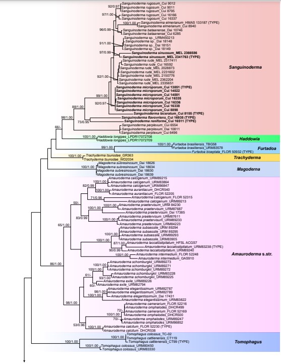
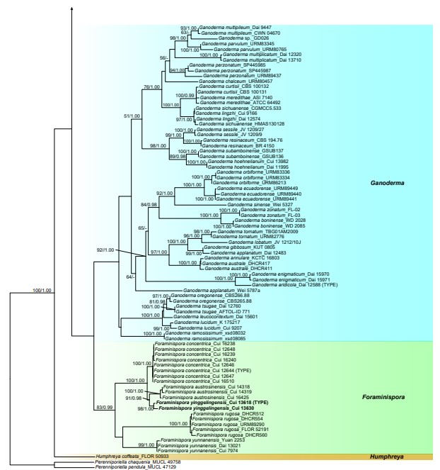
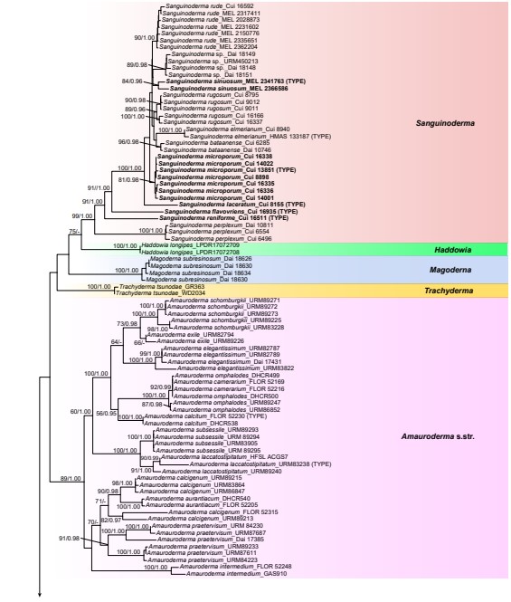
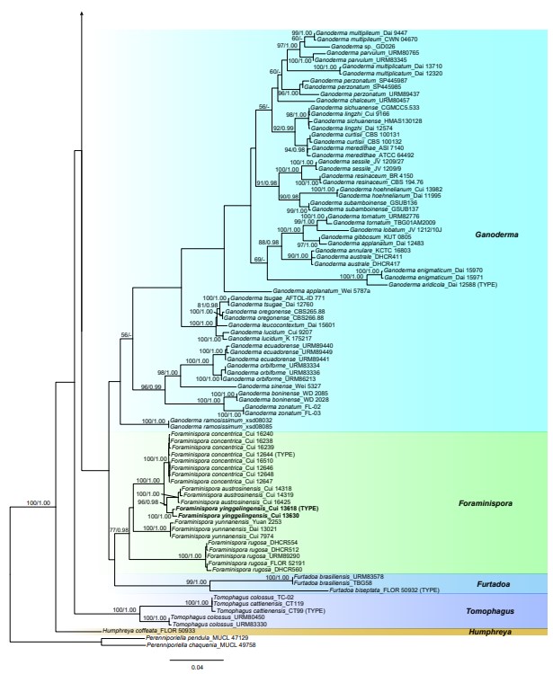
Notes: — All examined specimens in Sanguinoderma were found in the Paleotropics and in Oceania. In the 4-gene and 6-gene based phylogenetic analyses (Fig. 1, 2) these species nested in eleven highly supported lineages. Sanguinoderma presents similar macro- and micro-morphology to Amauro derma s.str.; however, it can be distinguished by its conspicuous reaction in the pore surface, which rapidly changes to blood red when bruised. With the addition of morphological evidence, five new species are described and five new combinations are proposed and re-described. One taxon with insufficient mor- phological characters to separate it from other taxa is treated as Sanguinoderma sp.

Fig. 1 ML analyses of Ganodermataceae based on dataset of ITS+nLSU+RPB1+TEF. ML bootstrap values higher than 50 % and Bayesian posterior proba- bilities values more than 0.95 are shown. New species are in bold.

Fig. 1 (cont.)

Fig. 2 ML analyses of Ganodermataceae based on dataset of ITS+nLSU+RPB1+RPB2+TEF+TUB. ML bootstrap values higher than 50 % and Bayesian posterior probabilities values more than 0.95 are shown. New species are in bold.

Fig. 2 (cont.)
Species
