Salinomyces polonicus Czachura & Piątek, in Czachura, Owczarek-Kościelniak & Piątek, Fungal Biology 125(6): 463 (2021)
Index Fungorum Number: IF 838511, MycoBank Number: MB 838511, Facesoffungi Number: FoF 15857
Etymology – The name refers to Poland, where the samples were collected and the fungal strains were isolated.
On MEA: Mycelium consisting of hyaline (when young), brown (when mature), septate, branched, verrucose or smooth hyphae, 2e7 mm wide. Chlamydospores intercalary, globose, brown, one- or two-celled, 7.5e15.5 × 5.5e10.5 mm. Conidiophores and conidia absent. On PDA: Mycelium consisting of hyaline (when young), pale brown to brown (when mature), septate, branched, sometimes slightly verruculose hyphae, 2.0e6.5 mm wide. Chlamydospores rarely produced. Multi-cellular bodies, brown, 12.5e17 × 11.5e13.5 mm. Conidiogenous cells intercalary on hyphae. Conidia one- or two-celled, pale brown to brown, smooth or rugose, 5.5e9.5 × 4.5e7.5 mm.
Culture characteristics – Colony on MEA rounded, greenish gray, stiff and dense, with irregular surface, reaching 22.2 mm in diameter at 20 °C after 8 weeks growth. Reverse navy blue with lighter margin. Colony on PDA rounded, blackish gray, wrinkled, stiff and dense, with irregular surface and brown margin, reaching 21.3 mm in diameter at 20 °C after 8 weeks growth. Reverse dark navy blue.
Type – Poland, Silesian Province, Cieszyn County: De˛bowiec-graduation tower, isolated from brine, 12 Nov. 2017, P. Czachura (holotype: KRAM F-59684; culture ex type: CBS 147480.
GenBank Accession Numbers – CBS 147480 – ITS: MW581542, LSU: MW581538, rpb2: MW583643; CBS 147481 – ITS: MW581543, LSU: MW581539, rpb2: MW583644
Other material examined – Poland, Silesian Province, Cieszyn County: De˛bowiec-graduation tower, isolated from brine, 12 Nov. 2017, P. Czachura (culture: CBS 147481).
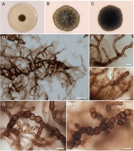
Figure 1 – Salinomyces polonicus on MEA. (AeC) Upper side and reverse of colony, respectively; (D) mycelium containing hyphae and chlamydospores; (EeF) pigmented (mature) and hyaline (young) hyphae; (GeH) chlamydospores. Scale bars: D = 50 mm, E-H = 10 mm.
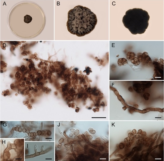
Figure 2 – Salinomyces polonicus on PDA. (AeC) Upper side and reverse of colony, respectively; (D) mycelium containing hyphae and multi-cellular bodies; (E) multi-cellular bodies; (F) pigmented hypha; (GeI) hyphae with conidiogenous cells; (JeK) conidia. Scale bars: D = 50 mm, EeK = 10 mm.