Russula shoreae D. Chakr., A. Ghosh, K. Das & Buyck sp. nov.
MycoBank number: MB; Index Fungorum number: IF; Facesoffungi number: FoF 11435;
Description
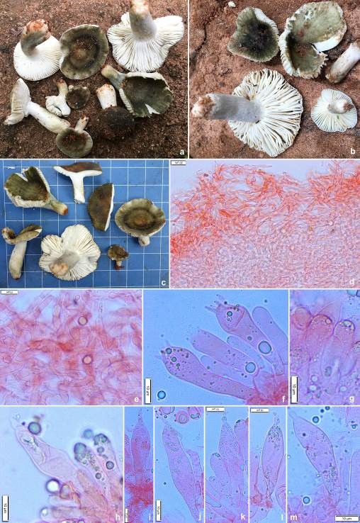
Pileus small to medium-sized, 12–70 mm in diam., convex when young, becoming planoconvex to applanate, uplifted with age, centrally depressed to umbilicate with maturity; margin decurved to plane with age, entire; cuticle viscid and shiny when moist, dull upon drying, peeling to 1/2nd of the radius, dark green (27F5–6) to dull green (26C–D3–4) at young stage with paler margin (26D4), with maturity dark green (27F5–6) at centre with dark green concentric rings (25–26F7–8) in combination of greyish green (26D3–4). Pileus context up to 6 mm thick at the disc, thinning towards the margin, compact, brittle, chalky white (1–2A1), unchanging after bruising or cutting; turning salmon pink (6A4) with FeSO4 and deep to dark turquoise (24E–F7–8) in guaiacol. Lamellae up to 4 mm deep, narrowly adnate to adnexed, subdistant to close to (9–13/cm at pileus margin), chalky white (1–2A1), forked near the stipe apex, midway to the margin, or near the margin; lamellulae present in different lengths; edges entire and concolorous. Stipe 10–67 × 5–22 mm, cylindrical to clavate, central, firm and brittle; surface dry, smooth, chalky white (1–2A1) with dull green (26D4) tinges. Stipe context solid when young, becoming stuffed to hollow with maturity, chalky white (1–2A1), unchanging after bruising or cutting; salmon pink (6A4) with FeSO4 and deep to dark turquoise (24E–F7–8) in guaiacol. Odour not distinctive. Taste mild. Spore print not obtained.
Basidiospores sub-globose, broadly ellipsoid to ellipsoid, rarely globose, (5.5–)6.3–7.0–7.7(–8.4) × (4.8–)5.4–6.0–6.6(–7.8) μm, Q=(1.02–)1.09–1.17–1.25(–1.41); ornamentation composed of amyloid isolated warts (up to 0.5 μm high); warts pustulose or rounded, sometimes fused with each other; suprahilar spot distinct, large but inamyloid, apiculi up to 1.5 μm high. Basidia (46–)51–57–62(–65) × (9–)10–11–12(–13) μm, 4-spored, subclavate, tapering towards base, sterigmata up to 6 μm long. Hymenial cystidia near the lamellae sides (50–)53.3–60.7–68(–76) × (7–)9.8–11.6–13.5(–15.2) µm, rare, cylindrical, subclavate, clavate to fusiform with rostrate to moniliform apex, emergent up to 22 μm above the other elements of the hymenium; contents finely crystalline, without reaction in sulfovanillin. Lamellae edges fertile with basidia and cystidia. Hymenial cystidia near the lamellae edges usually smaller and narrower, measuring (46–)46.2–50.7–55.2(–55) × (9–)9.1–10.7–12.2(–12) µm, cylindrical to fusiform with rostrate to moniliform apex; contents finely crystalline, without reaction in sulfovanillin. Subhymenium layer 35–40 µm thick, pseudoparenchymatous. Hymenophoral trama mainly composed of large nests of sphaerocytes and intermixed with hyphal elements. Pileipellis orthochromatic in Cresyl blue, sharply delimited from the underlying sphaerocytes of the context, 276–307 μm deep, two layered; vaguely divided in 96–91 μm deep suprapellis of relatively dense, composed of erect or ascending hyphal terminations, arranged in a trichodermal structure and dispersed primordial hyphae; subpellis 180–216 μm deep, composed of more or less dense, horizontally oriented hyphae. Acidoresistant incrustations absent. Hyphal terminations near the pileus margin often slightly flexuous, thin-walled, composed of chain of 1–3 cells, branched at the subterminal cells or the cells just below; terminal cells measuring (11–)13.1–21–28.9(–44.5) × (3–)3.8–4.6–5.3(–7) μm, mainly subulate or cylindrical, apically acute and distinctly attenuated or obtuse-rounded; subterminal cells mainly cylindrical, but sometimes wider. Hyphal terminations near the pileus centre of similar structure, terminal cells slightly less wide, measuring (9–)12.4–19.4–26.3(–30.3) × (2.6–)3.1–3.8–4.5(–5) μm, mainly subulate or cylindrical, apically acute and distinctly attenuated or obtuse-rounded; subterminal cells mainly cylindrical, but sometimes wider or sometimes with lateral appendages. Pileocystidia near the pileus margin typically one celled, flexuous, thin-walled; terminal cells (14–)17.5–39.9–62.2(–96) × (2.4–)3.5–4.7–5.8(–6) μm, mainly subulate, apically mostly mucronate or with short appendages; contents finely crystalline, without reaction in sulfovanillin. Pileocystidia near the pileus centre one-celled, flexuous, thin-walled, slightly shorter; terminal cells (22–)23.9–31.7–39(–46) × (3.5–)3.8–4.3–4.7(–5) μm, mainly subulate, apically mostly mucronate or with short appendages; contents finely crystalline, without reaction in sulfovanillin. Clamp connections absent in all parts.
Material examined: India, West Bengal, Jhargram district, Lodhasuli, alt. 80 m, N 22°19’57” E 87°02’47” E, 27 August 2021, D. Chakraborty, NPDF917-10L (CAL 1864, holotype!).
Sequence data: ITS: OL461227 (nrITS Holotype) and OL461230 (nrITS) LSU: ON365930 (nrLSU, holotype), ON365931 (nrLSU) mtSSU: ON387509 (mtSSU, holotype), ON387514 (mtSSU) rpb2: ON398069 (rpb2, holotype), ON398070 (rpb2)
Notes: nBLAST results of the obtained sequence place R. shoreae in subg. Heterophyllidiae, which was also clearly suggested by its morphological characters, including the inamyloid suprahilar spot, the typical ramifying hyphal extremities at the pileus surface composed of chains of more or less inflated, short cells that become gradually narrower toward the terminal cell and one-celled, narrow pileocystidia.
In our ITS phylogenetic analysis, the here newly described R. shoreae is placed sister to the Chinese R. verrucospora Y. Song & L. H. Qiu. Both are again placed sister to the North American and equally green R. redolens Burl. These three species form a strongly supported clade, which is placed sister, again with strong statistical support, to the annulate R. brunneoannulata Buyck of the African subsect. Aureotactinae Heim ex Buyck (Buyck 1994). All these species have very similar microscopic features of pileipellis and share the same type of spore ornamentation consisting of isolated blunt warts. Some of the abovementioned species were also grouped with strong support in recent multilocus phylogenies. A representative sampling of species belonging to subg. Heterophyllidiae was distributed over four significantly supported clades in the combined multilocus phylogeny, based on 28S, rpb2 and tef1 loci, that was published by Wang et al. (2019). In that phylogeny, subsect. Substriatinae was introduced as a new subsection that grouped with Aureotactinae as one of the four strongly supported clades in the subgenus. This topology was never recovered in ITS-based phylogenies, nor in the combined multilocus (based on the same loci) published by Vera et al. (2021) where the placement of Aureotactinae was affected by the introduction of R. redolens and no longer grouped with Substriatinae.

Figures 1. Russula shoreae (CAL 1864, holotype) a–c Fresh and dissected basidiomata in the field and basecamp. d, e Transverse section through pileipellis showing elements. f Transverse section through lamellae showing basidia. g, h Transverse section through lamellae showing hymenial cystidia near the lamellae edges. i−m Transverse section through lamellae showing hymenial cystidia near the lamellae sides. Scale bars: d = 20 μm, e−m = 10 μm.
