Robillarda africana Crous & Giraldo López, in Crous et al., IMA Fungus 6(1): 184 (2015)
MycoBank number: MB 812797; Index Fungorum number: IF 812797; Facesoffungi number: FoF 10700;
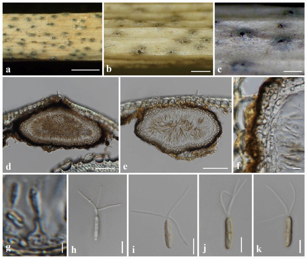
Saprobic on grass litter. Sexual morph: Undetermined. Asexual morph: Conidiomata 100–140 × 170–230 µm (x̄ = 125 × 190 μm, n = 20), pycnidial, scattered, globose to subglobose, semi immersed, unilocular, dark brown to black, ostiolate. Conidiomatal wall 30–40 µm, brown to dark brown, arranged in a textura angularis, cells towards the inside hyaline, at the outside darker, fusing and indistinguishable from the host tissues. Conidiophores reduced to conidiogenous cells. Conidiogenous cells 4–5 × 2–3 µm (x̄ = 4.5 × 2.5 μm, n = 20), phialidic, ampulliform to subcylindrical, hyaline, smooth, guttulate, deliquescing at maturity. Conidia 10–12 × 1.5–3 µm (x̄ = 11 × 2.2 μm, n = 30), cylindrical to fusiform, straight or slightly curved, 1-septate, slightly constricted at median septum, hyaline to pale brown, rounded at both ends, smooth-walled, 3 extracellular appendages, 11–16 µm long, arising subapically of the conidial sheath.
Material examined – THAILAND, Chiang Mai Province, Mae Teang District, Mushroom Research Center (M.R.C.), on dead culms of unidentified grass (Poaceae), 18 March 2016, Ishani D. Goonasekara, IGm25 (new host and geographical record), living culture MFLUCC 16-0888.
Distribution – South Africa, Thailand (Crous et al. 2015, Gooansekara et al. 2022)
Sequence data – MFLUCC 16-0888: ITS: OL824826 (ITS5/ITS4); LSU: OL824828 (LROR/LR5)
Notes – As morphological characters examined largely overlap with those of Robillarda africana (CBS 122.75), we therefore report our collection (MFLUCC 16-0888) as a new host and geographical record of R. africana from dead grass in Thailand. Robillarda africana (CBS 122.75) was initially introduced by Crous et al. (2015) from South Africa (exact location and substrate unknown). Both isolates (CBS 122.75 and MFLUCC 16-0888) have pycnidial, globose to subglobose, semi immersed, unilocular conidiomata, ampulliform to subcylindrical, hyaline conidiogenous cells (4–5 × 2–3 µm vs 3–10 × 2–4 µm) and cylindrical to fusiform, straight or slightly curved, 1-septate, hyaline to pale brown conidia (10–12 × 1.5–3 µm vs 11–12 × 2.5–3 µm) with extracellular appendages (Crous et al. 2015).

Fig. Robillarda africana (IGm25, new host and geographical record): a, b. Appearance of conidiomata on host. c. Close-up of conidioma. d, e. Sections of conidiomata. f. Conidiomatal wall. g. Conidiogenous cells and developing conidia. h–k. Conidia. Scale bars: d, e = 50 µm, f = 10 µm, g = 2 µm, h–k = 5 µm.
