Rhexoacrodictys melanospora S.X. Bao, R.J. Xu & Qi Zhao, sp. nov.
MycoBank number: MB559983; Index Fungorum number: IF559983; Facesoffungi number: FoF 12898
Etymology – The specific epithet ‘melanospora’ refers to the dark conidia of the fungus
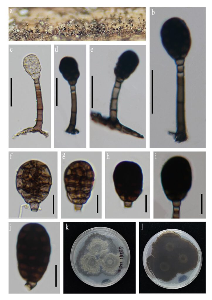
Saprobic on decaying woods in terrestrial habitat. Asexual morph: Conidiophores 19–60 × 3–5 μm (Q = 40 × 4 μm, n = 20), macronematous, mononematous, erect, straight or somewhat fexuous, pale brown to brown, cylindrical, thick-walled, smooth, 2–4 septate. Conidiogenous cells 2–6 × 3–4 μm (Q = 4 × 3.5 μm, n = 10), monoblastic, integrated, terminal, pale brown, cylindrical, with 1-3 pale brown percurrent extensions. Conidia 16–32 × 12–21 μm (Q = 23 × 16 μm, n = 40), solitary, acrogenous, ellipsoidal to obovoid or somewhat subglobose, muriform, with 2–5 transverse and several oblique septa, pale brown when immature, becoming dark brown at maturity, thick and smooth-walled. Sexual morph: Undetermined.
Culture characteristics: Colonies on PDA, reaching 4 cm diam. after 15 days at room temperature, colonies irregular, surface taupe, mycelium dense, and reverse blackish.
Material examined: —China, Yunnan Province, Fugong Country, decayed wood in terrestrial habitats, 13 May 2021, Song Wang, GLG41-1 (HKAS 124580, holotype), ex-type living culture KUNCC 22-12406; ibid., Derung-Nu Autonomous Country of Gongshan, 15 May 2021, Song Wang, GLG41-2 (HKAS 124581, paratype), ex-type living culture KUNCC 22-12411.
Notes: Morphologically, it fits well the generic concept of Rhexoacrodictys in having rhexolytically seceding conidia (Baker et al. 2002; Zhao et al. 2010). Among the 5 species epithets of Rhexoacrodictys (Index Fungorum 2022), two species R. broussonetiae and R. fuliginosa have no molecular data available, the other three species can be distinct from R. melanospora by molecular data (Fig. 1). Although their molecular data deficiency, R. melanospora can be distinguished from R. broussonetiae and R. fuliginosa that it has shorter conidiophores (19–60 μm in R. melanospora vs. 70–90 μm in R. broussonetiae vs. up to 85 μm in R. fuliginosa) and longer conidia (16–32 μm in R. melanospora vs. 17–28μm in R. broussonetiae vs. 17–27 μm in .
We therefore introduce R. melanospora as new to the genus. A morphological comparison of Rhexoacrodictys species is summarized in Table 2 and figure plate of R. melanospora is illustrated in Figure 2.

FIGURE 2. Rhexoacrodictys melanospora (a) Colonies on natural substrate. (b–e) Conidiophores, conidiogenous cells and conidia. (f–j) Conidia (k–l) Colony on PDA (surface and reverse). Scale bars b–e = 20 μm f–j = 10 μm,
