Hymenotorrendiella indonesiana Crous & P.R. Johnst., in Crous et al., Persoonia 44: 349 (2020)
MycoBank number: MB 835414; Index Fungorum number: IF 835414; Facesoffungi number: FoF 12899;
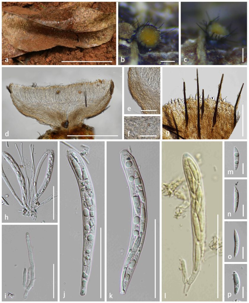
Saprobic on leaf litters on Eucalyptus sp. Sexual morph: Apothecia 0.4–0.8 mm wide × 0.5–1.0 mm high (x̅ = 0.6 × 0.7 mm, n = 20), superficial, scattered or gregarious in groups, stipitate when dry. Disc flat to slightly concave when dry, smooth, yellowish central with off-white edge. Receptacle cupulate, concolorous at the edge of the disc, covered with dark brown setae. Stipe 0.2–0.4 mm (x̅ = 0.3 mm, n = 20), short, smooth, dark brown. Setae mostly 15–25 per apothecium, 180–270 μm long, smooth, dark brown but paler on apex, 7–8-septate, thick-walled, constricted at the base and connected with the central ectal excipulum cell. Hymenium 100–140 μm, hyaline with yellowish brown contents. Ectal excipulum 66–74 μm thick, comprised of 3–4 layers, thin-walled, hyaline cells of textura angularis with yellowish or brown exudate, inner layer of ectal excipulum comprised of narrow textura prismatica cells at stipe, non-gelatinous. Medullary excipulum 57–63 μm thick, comprised of thin-walled, loosely hyphae of textura intricata, 3.2–4.0 μm wide, hyaline, non-gelatinous. Paraphyses 2.8–3.4 μm (x̅ = 3.3 μm, n = 30) in the widest, sometimes branched and septate near the base, rounded apex, hyaline. Asci 88–110 × 7–10 μm (x̅ = 99 × 9 μm, n = 40), 8-spored, cylindric to subclavate, subconical apex with amyloid apical pore in Melzer’s reagent, tapering to subtruncate base. Ascospores (17.5–)18.0–26.0(–28.0) × (3.7–)4.0–4.9 μm (x̅ = 22 × 4.4 μm, n = 100), Q = (3.8–)4.1–6.2(–7.1) μm, Qm = 5.0 ± 0.6 μm, overlapping uni- to bi-seriate, fusiform with umbrella-shaped, mucilaginous appendage at the ends, slightly curved, hyaline, thin-walled, smooth, aseptate with unipolar or bipolar and irregular guttules. Asexual morph: Undetermined.
Material examined – China, Yunnan Province, Puer City, Jingdong County, dead leaf litter of Eucalyptus sp., 9 June 2022, Cuijinyi Li LCJY-772 (HKAS 124584).
GenBank accession numbers – ITS: OP321584.
Known distribution (based on molecular data) – Indonesia, Malaysia (Crous et al. 2006b, 2020), China (this study).
Known hosts (based on molecular data) – Eucalyptus urophylla, Eucalyptus sp. (Crous et al. 2006b, 2020, this study).
Notes – Our collection is found in the subtropical evergreen broad-leaved forest and shares similar morphological features with Hymenotorrendiella indonesiana (CPC 11050) by having small cupulate apothecia, distinguishable coloured stipe from the receptacle, dark and long setae arising from the central cells of ectal excipulum and fusiform ascospores with umbrella-shaped mucilaginous appendages at both ends (Crous et al. 2006b). The comparison of the base pairs between our strain and CPC 11050 shows a 0.85% difference in the ITS region (7/823). Based on the phylogenetic analyses of ITS gene fragment (Fig. 43), our collection clustered with Hymenotorrendiella indonesiana (CPC 11049 and CPC 11050) with 96% maximum likelihood bootstrap support, 86% maximum parsimony bootstrap support and 0.99 Bayesian probability statistical supports, respectively. This is the first record of this species in China.

Fig. 1– Hymenotorrendiella indonesiana (HKAS XXX, new geographical record). a Fresh ascomata on the leaf. b, c Dried ascomata on the leaf. d Vertical section of ascoma. e Ectal excipulum. f Medullary excipulum. g Hairs. h Asci and paraphyses. i Paraphyses j–l Asci (l Ascus in Meltzer’s reagent). m–p Ascospores. Scale bar: a = 4 cm, b–d = 300 μm, e = 70 μm, f = 40 μm, g = 100 μm, h = 50 μm, i = 30 μm, j–l = 50 μm, m, n–p = 10 μm.
