Quasiramularia phakopsoricola I-Chin Wei & R. Kirschner, sp. nov. Figures 2–5, 7–8
MycoBank number: MB 558772; Index Fungorum number: IF 558772; Facesoffungi number: FoF;
Etymology: Referring to the genus of the host fungus, Phakopsora.
Hyphae and conidiophores formed in white to dirty white dusty-dry pustules on rust uredinia on the abaxial side of living leaves. Conidiophores emerging from uredinia, loose, erect to decumbent, effuse, more or less straight, subcyclindric, septate, hyaline or subhyaline, smooth to finely verruculose, simple or with few short branches, (13–)22–44(–60) × 2–3(–4) µm (n = 30). Conidiogenous cells mostly terminal, rarely subterminal or lateral, subcyclindric or slightly swollen, (4–)10–22(–25) × 2–3 µm (n = 30); conidiogenous loci thickened and slightly darkened. Conidia catenate, ellipsoid-ovoid or subcylindric-fusiform, ends rounded to somewhat pointed, (6–)7–15(–20) × 4–5(–7) µm (n = 30), hyaline or subhyaline, smooth to rough, hila thickened, slightly darkened, less than 1-μm diam.
Colonies growing slowly on CMA at room temperature, reaching approx. 25-mm diam. after 1 month, whitish, subeffuse to powdery on top side, dark greenish pigment released on reverse side. Conidiophores similar to those on the host, (42–)49–131(–150) × 2 μm (n = 30). Conidiogenous cells subcyclindric, 5–19(–22) × 2 µm. Conidia catenate, ellipsoid-ovoid, fusiform, or subcylindric-fusiform, ends rounded to somewhat pointed, (7–)10–18(–23) × 3–5(–6) µm (n = 30), hyaline, smooth to rough, hila thickened, slightly darkened. Specimens examined (dried specimens on natural substrate, if not stated otherwise): On Phakopsora ampelopsidis Dietel & P. Syd. (Phakopsoraceae) on Ampelopsis brevipedunculata (Vitaceae), Taiwan, Taoyuan City, Zhongli District, north of National Central University, Neicuo 3rd Rd., 24.97510107, 121.19031800, ca. 135 m, 10. Nov 2015, I.C. Wei & R. Kirschner WIC009 (TNM, holotype), ex-type strain BCRC FU30724, ITS MT741740, SSU MT741743, LSU MT741723; ibid., 04. Dec 2015, I.C. Wei WIC018 (TNM); ibid., 28. Dec 2015, I.C. Wei WIC019A (on leaves) and WIC019B (dried culture) (TNM); ibid., 20. Mar 2017, R. Kirschner 4383 (TNM); New Taipei City, Yingge District, Yingge Rock hiking trail, ca. 24.958814, 121.360379, ca. 80 m, 23. May 2017, R. Kirschner 4400 (TNM); ibid., 27. Oct 2018, R. Kirschner 4689 (TNM); Taipei City, Zhongshan District, Jian Butterfly Garden, 25.086900, 121.554600, ca. 350 m, 01. Feb 2017, R. Kirschner 4365 (TNM), strain BCRC FU30777, ITS MT741738, SSU MT741741, LSU MT741721, RPB2 LC535131. On Ph. ampelopsidis on Parthenocissus tricuspidata (Siebold & Zucc.) Planch. (Vitaceae), Taipei City, Daan District, border of Fuzhoushan Park at Wolong Street, ca. 25.017834, 121.552391, ca. 10 m, 31. Mar 2019, R. Kirschner 4704 (TNM), ITS MT741739, SSU MT741742, LSU MT741722, RPB2 LC535132, ibid., 19. Aug 2021, R. Kirschner 5313 (TNM).
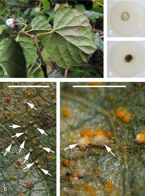
Fig. 2 Quasiramularia phakopsoricola. a White spots formed on orange rust sori (Phakopsoraceae) on the abaxial side of a leaf of Ampelopsis brevipedunculata (R. Kirschner 4400). b, c Whitish pustules formed by conidiophores and conidia (arrows) on the orange rust sori on Parthenocissus tricuspidata in situ (R. Kirschner 5313). d Petri dish (9-cm diam.) with culture (WIC009) seen from above (CMA, ca. 1 month). e Same culture as in d, reverse. Scale bars: b = 2 mm, c = 1 mm
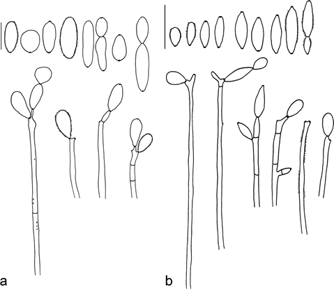
Fig. 3 Quasiramularia phakopsoricola, light microscopy. a Conidiophores and conidia from the host in situ. b Conidiophores and conidia formed by the fungus in culture (WIC009). Scale bars = 10 µm
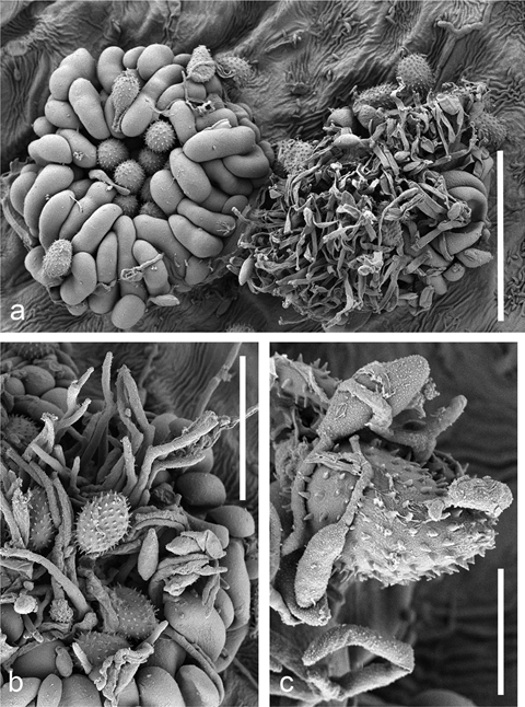
Fig. 4 Quasiramularia phakopsoricola (WIC009) on a rust fungus (Phakopsoraceae) on Ampelopsis brevipedunculata as seen by scanning electron microscopy (SEM). a Two rust sori, the right one heavily overgrown by Q. phakopsoricola. b Conidiophores among urediniospores and paraphyses. c Conidia of Q. phakopsoricola germinating on the surface of a urediniospore. Scale bars: a = 50 µm, b = 30 µm, c = 10 µm
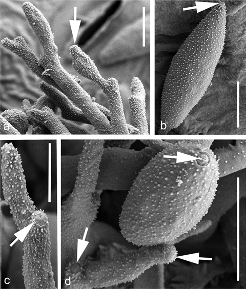
Fig. 5 SEM photographs show- ing conidiogenous cells and conidia with surface ornament, conidiogenous loci, and hila of conidia of Q. phakopsoricola (WIC009). a Conidiogenous cells with terminal geniculations and conidiogenous loci (one locus indicated by an arrow). b Fusiform conidium with hilum (arrow). c Conidiogenous cell with conidiogenous locus and a central pore (arrow). d Conid- iogenous loci and one hilum of a conidium (arrows). Scale bars: a, b, d = 5 µm, c = 3 µm
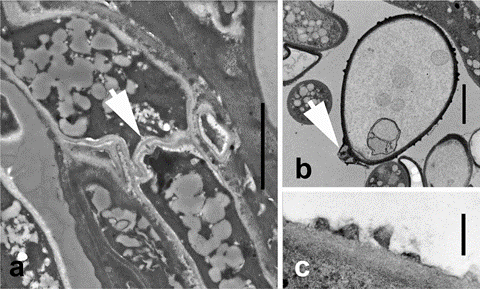
Fig. 7 TEM photographs of cell wall structures of Q. phakopsoricola. a Septal pore (arrow) in a hypha from material in nature (WIC009). b Conidium with surface ornament. The arrow indicates the hilum as shown in Fig. 5d (R. Kirschner 4689). c Surface ornament at higher magnification. Scale bars a = 2 μm, b = 2 μm, c = 0.2 μm
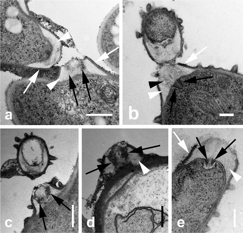
Fig. 8 TEM photographs of conidiogenous loci of Q. phakopsoricola (R. Kirschner 4689). a Detachment of conidium base from a conidiogenous locus. Cell walls of both structures have thickened electron- transparent material (white arrowheads) between the thin electron- dense outer layer of cell wall (white arrows) which is connected to the cell wall of the entire conidiogenous cell. Note the more electron-dense (slightly shaded) presumably ring-shaped structure (black arrows) embedded in the electron-transparent material (arrows). b Conidiogenous locus with shaded area (arrowhead) embedded in electron-transparent material (white arrowhead) and extending into a presumably ring-shaped electron-dense structure (black arrows) which is projecting into the cytoplasm. c, d Strongly electron-dense presumably ring-shaped structures (black arrows) embedded in electron-transparent material (white arrowhead in d) within conidiogenous locus. e Presumably ring-shaped electron-dense structure (black arrows) appearing to be pressed by copious electron-transparent material (white arrowhead) into the cytoplasm. An electron-dense outer cell wall layer (white arrow) is continuous with the outer cell wall layer of the conidiogenous cell. Scale bars: a, c, d = 0.5 μm; b, e = 0.2 μm
