Pseudoroussoella elaeicola (Konta & K.D. Hyde) Mapook & K.D. Hyde, comb. nov.
MycoBank number: MB 557352; Index Fungorum number: IF 557352; Facesoffungi number: FoF 07819; Fig. 78
≡ Roussoella elaeicola Konta & K.D. Hyde, in Phookam- sak et al., Fungal Divers (2019)
Holotype: THAILAND, Chiang Rai Province, on dead petiole of Elaeis guineensis (Arecaceae), 25 November 2014, S. Konta, HR02d (MFLU 15-0022), ex-type culture, MFLUCC 15-0276
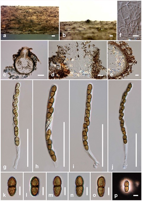
Saprobic on dead stems of Chromolaena odorata. Sexual morph: Ascomata 225–475 µm high × 240–400 µm diam. ( x̄ = 365 × 325 µm, n = 5), immersed, solitary, appearing as dark spots, coriaceous, subglobose, dark brown to black. Ostiolar neck protruding. Peridium (20–)30–50 µm wide, several layers, inner layers comprising hyaline to light brown cells of textura epidermoidea, outer layers comprising brown to dark brown cells of textura intricata. Hamathecium com- prising 1–2 µm wide, cylindrical to filiform, septate, trabeculate pseudoparaphyses. Asci (70–)95–135 × 6–8.5 µm ( x̄ = 108 × 7 µm, n = 10), 8-spored, bitunicate, cylindrical to clavate, straight or slightly curved, apically rounded, short pedicellate with small ocular chamber. Ascospores 10–14 × 4.5–6 µm (x̄ = 12.5 × 5.5 µm, n = 25), uniseriate, initially hyaline to pale brown, septate when immature, becoming yellowish brown at maturity, oval to ellipsoid, 1-septate, constricted at the septum, straight or slightly curved, slightly widest at the upper cell and tapering towards obtuse ends, with a reticulate spore wall ornamentation, surrounded by hyaline gelatinous sheath observed clearly when mounted in Indian ink. Asexual morph: Undetermined.
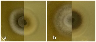
Culture characteristics: Ascospores germinating on MEA within 48 h. at room temperature and germ tubes produced from both ends. Colonies on MEA circular, mycelium slightly raised, velvety with moderately fluffy, filamentous, cultures white, grayish-brown from the centre of the colony with white at margin on surface, brown to pale olivaceous- brown in reverse from the centre of the colony, white to creamy-white at margin (Fig. 79a).
Pre-screening for antimicrobial activity: Pseudoroussoella elaeicola (MFLUCC 17-1483) showed antimicrobial activity against E. coli with a 10 mm inhibition zone when compared to the positive control (9 mm), but no inhibition of B. subtilis and M. plumbeus.
Known hosts and distribution: On dead petiole of Elaeis guineensis (Arecaceae) in Thailand (Phookamsak et al. 2019).
Material examined: THAILAND, Lampang Province, Ngao, on dead stems of Chromolaena odorata, 21 September 2016, A. Mapook (LP5, MFLU 20-0357); living culture MFLUCC 17-1483 (new host record).
GenBank numbers: LSU: MT214442, ITS: MT214348, SSU: MT214396, TEF1: MT235772, RPB2: MT235808
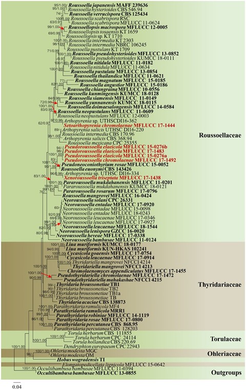
Notes: A phylogenetic analyses show that the strain MFLUCC 17-1483 grouped with Pseudoroussoella elaeicola (= Roussoella elaeicola) (Fig. 76). In a BLASTn search of NCBI GenBank, the closest match of the ITS sequence of our strains with 99.80% similarity was Roussoella sp. (strain MFLUCC 17-2059, MH744730). The closest match with the LSU sequence with 98.47% similarity was Arthopyrenia salicis (strain MUT < ITA > :4879, KP671722). The closest match with the SSU sequence with 93.95% similarity was Roussoella intermedia (strain KT 2303, AB524483). The closest match with the TEF1 sequence with 96.06% similarity was Arthopyrenia sp. (strain UTHSC: DI16-334, LT797127), while the closest match with the RPB2 sequence with 90.92% similarity was R. euonymi (strain CBS 143426, MH108007). We therefore, identify our isolates as P. elaeicola based on phylogenetic analyses with morphological comparison (Table 17). In this study, we isolated P. elaeicola from Chromolaena odorata collected in Thailand, and the isolate is introduced here as a new host record.
Table 17 Synopsis of sexual morph species with similar morphological features discussed in this study
| Species | Ascomata (µm) | Peridium (µm) | Asci (µm) | Ascospores (µm) | References |
| Pseudoroussoella elaeicola(= Roussoella elaeicola, MFLUCC 15-0276)
|
315–410 high, 325–350 diam. | 25–70 | 70–140 × 6–9 | 10–15 × 3–6, 1-septate | Phookam- sak et al. (2019) |
| P. elaeicola (MFLUCC 17-1483, LP5)
|
225–475 high × 240–400
diam. |
(20–)30–50 | (70–)95–135 × 6–8.5 | 10–14 × 4.5–6, 1-septate | This study |
| Xenoroussoella triseptata
(MFLUCC 17-1438, DP22)
|
(155–)170–220 high × 185–
235 diam. |
10–15 | 55–70 × 10.5–15 | 13.5–17 × 5–7.5, 3-septate | This study |

Fig. 76 Phylogram generated from maximum likelihood analysis based on combined dataset of LSU, ITS, TEF1, RPB2 and SSU sequence data. Eighty- four strains are included in the combined sequence analysis, which comprise 4416 characters with gaps. Tree topology of the ML analysis was similar to the BYPP. The best scoring RAxML tree with a final likelihood value of − 35074.673502 is presented. The matrix had 1690 distinct alignment patterns, with 39.45% of undetermined characters or gaps. Estimated base frequencies were as follows: A = 0.246392, C = 0.254324, G = 0.268438, T = 0.230846; substitution rates: AC = 1.657341, AG = 4.989387, AT = 2.205689, CG = 1.285998, CT = 10.615734, GT = 1.000000; gamma distribution shape parameter α = 0.486933. Bootstrap sup- port values for ML equal to or greater than 60% and BYPP equal to or greater than 0.90 are given above or below the nodes. Newly generated sequences and new combination are in dark red bold and type species are in bold. Occultibambusa bambusae (MFLUCC 11-0394) and O. bambusae (MFLUCC 13-0855) are used as outgroup taxa

Fig. 78 Pseudoroussoella elaeicola (new host record) a, b Appearance of immersed ascomata on substrate. c Section through ascoma. d Ostiole. e Peridium. f Pseudoparaphyses. g–j Asci. k–o Ascospores. p Ascospores with gelatinous sheath in Indian ink. Scale bars: a = 500 µm, b = 200 µm, c = 100 µm, g–j = 50 µm, d, e = 20 µm, f = 10 µm, k–p = 5 µm

Fig. 79 Culture characteristics on MEA: a Pseudoroussoella elaeicola (MFLUCC 17-1483). b Pseudoroussoella chromolaenae (MFLUCC 17-1492)
