Pseudopalawania siamensis Mapook and K.D. Hyde, sp. nov.
MycoBank number: MB 834935; Index Fungorum number: IF 834935; Facesoffungi number: FoF 11970; Figure 3
Etymology: Named after the country from where the fungus was collected, using the former name of Siam.
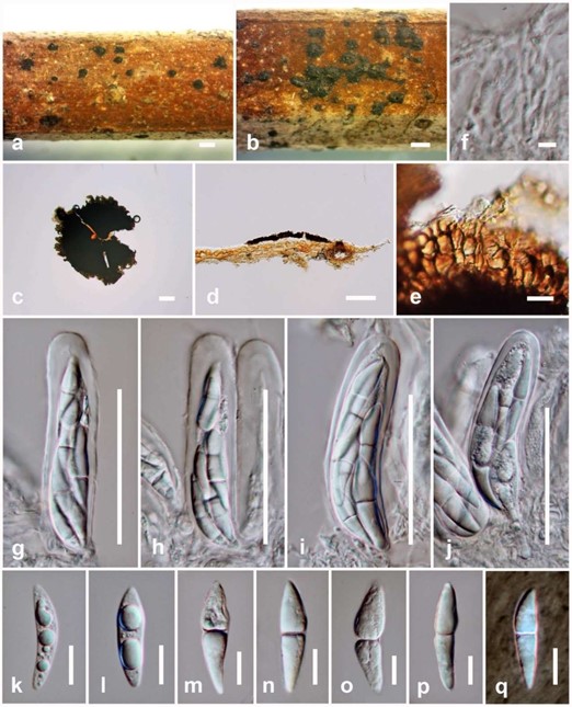
Saprobic on dead rachis of Caryota sp. Sexual morph: Ascomata 29–40 µm high × 270–290(–315) µm diam. (x = 32.5 × 292 µm, n = 5), superficial, solitary or scattered, sub-carbonaceous to carbonaceous, appearing as circular, flattened, dark brown to black spots, covering the host, without a subiculum, with a poorly developed basal layer and an irregular margin. Ostioles central. Peridium 10–20 µm wide, comprising dark brown or black to reddish-brown cells of textura epidermoidea to textura angularis. Hamathecium comprising 1–2.5 µm wide, cylindrical to filiform, septate, hyaline, branching pseudoparaphyses. Asci 65–85 × 15–21 µm (x = 75 × 18 µm, n = 10), eight-spored, bitunicate, fissitunicate, cylindric-clavate, straight or slightly curved, with an ocular chamber observed clearly when immature. Ascospores 25–37 × 5–11 µm (x = 29 × 7 µm, n = 20), overlapping, 2–3-seriate, broadly fusiform to inequilateral, pointed ends, hyaline, 1-septate, constricted at the septum, guttulate when immature, surrounded by hyaline and thin layers of gelatinous sheath, observed clearly when mounted in Indian ink. Asexual morph: Undetermined.
Culture characteristics: Ascospores germinating on MEA within 24 hrs. at room temperature and germ tubes produced from the apex. Colonies on MEA circular, slightly raised, filamentous, mycelium white at the surface and initially creamy-white to pale brown in reverse, becoming dark brown from the centre of the colony with creamy-white at the margin.
Pre-screening for antimicrobial activity: Pseudopalawania siamensis (MFLUCC 17-1476) showed antimicrobial activity against B. subtilis with a 16 mm inhibition zone and against M. plumbeus with a 17 mm inhibition zone, observable as full inhibition, when compared to the positive control (26 mm and 17 mm, respectively), but no inhibition of E. coli.
Material examined: THAILAND, Nan Province, on dead rachis of Caryota sp. (Arecaceae), 23 September 2016, A. Mapook (MFLU 20-0353, holotype); ex-type culture MFLUCC 17-1476.
Notes: Pseudopalawania is similar to Palawania in its superficial and flattened ascomata, with hyaline, 1-septate ascospores, but differs in its peridium wall patterns, shape of asci (cylindric-clavate vs. inequilateral to ovoid) with an ocular chamber and shape of ascospores (broadly fusiform to inequilateral vs. oblong to broadly fusiform) with a thin layer of gelatinous sheath. The gelatinous sheath in Palawania is thicker [24]. Pseudopalawania is also similar to Muyocopron in its superficial, flattened ascomata with similar peridium wall patterns, and asci with an ocular chamber; but differs in its sub-carbonaceous to carbonaceous ascomata, shape of asci and ascospores with surrounded by hyaline gelatinous sheath, 1-septate, while Muyocopron have coriaceous ascomata, aseptate ascospores with granular appearance and without gelatinous sheath [23]. In addition, the genus was compared with genera in Microthyriaceae of which no DNA sequence data are available, but the holotype specimens were re-examined in previous studies with morphological descriptions and illustrations [94–99], and neither of them matched our new fungus. Therefore, we introduce Pseudopalawania as a new genus with a new species P. siamensis from Thailand. The fungus is placed in Muyocopronaceae (Muyocopronales) with evidence from morphology and phylogeny.

Figure 3. Pseudopalawania siamensis (holotype) (a,b) Appearance of ascomata on substrate. (c) Squash mounts showing ascomata. (d) Section of ascoma. (e) Peridium. (f) Pseudoparaphyses. (g–j) asci. (k–p) Ascospores. (q) Ascospores in Indian ink. Scale bars: a, b = 500 µm, c, d = 100 µm, g–j = 50 µm, e, k–q = 10 µm, f = 5 µm.
