Babosia D.G. Knapp, Zagyva, Trappe & Kovács
MycoBank number: MB 834978; Index Fungorum number: IF 834978; Facesoffungi number: FoF14393;
Type: Babosia variospora, sp. nov. (described below).
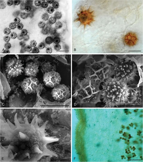
Diagnosis: Ascomata gasteroid, hypogeous. Differs from the phylogenetically most closely related Temperantia, Hydnobolites, and Stouffera by the dark sporogenous zone and dark brown spore color at maturity, the clear peridermal layer at maturity, and the variability of the ornamentation of the ascospores.
Etymology: Babosia, in honor and memory of the outstanding Hungarian mycologist Margit Babos (1931–2009).
Distribution: Central Europe (Hungary); one species known.

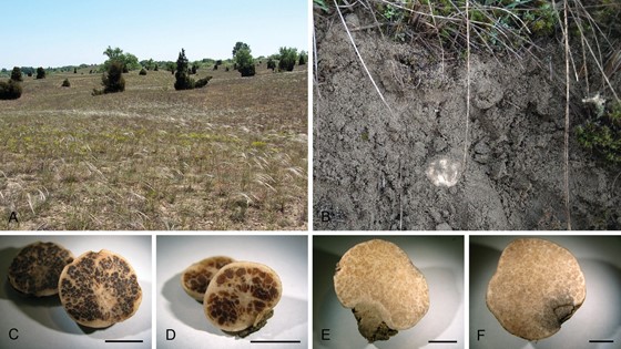
Figure 1. Sampling site and ascomata of the truffles described in the paper. A. The main collection site, a sandy grassland near Fülöpháza. B. Ascomata of Stouffera gilkeyae in sandy soil. C. Ascomata of Babosia variospora (KIPD_01). D. Ascomata of B. variospora (KIPD_02). E. Ascomata of S. gilkeyae (KIPD_05). F. Damaged part of the ascomata of the holotype KIPD_07 showing gray discoloration. Bars: C–F = 1 cm.

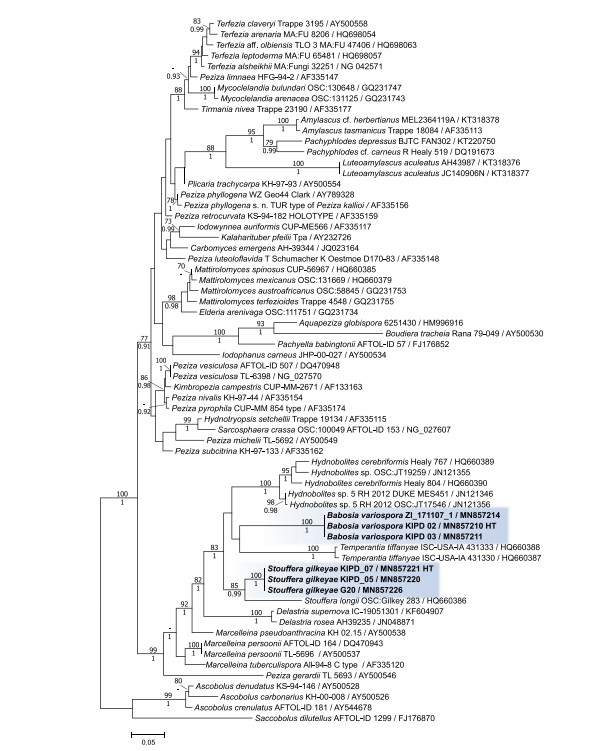
Figure 2. ML phylogenetic tree of 28S sequences of the two novel species and representative taxa of Pezizaceae. Bayesian posterior probabilities ≥0.90 are shown below branches; ML bootstrap support values ≥70% are shown above branches. Names of tips are followed by strain numbers and GenBank accession numbers separated by slashes. Sequences derived from holotype materials of the fungi introduced here are indicated as HT. The scale bar indicates 0.5 expected changes per site per branch.

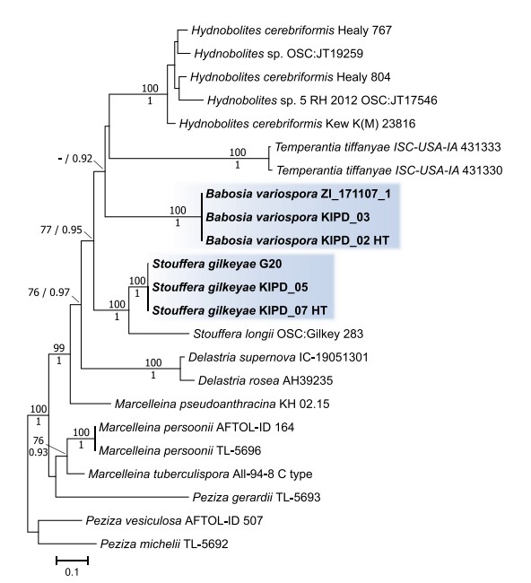
Figure 3. ML phylogenetic tree of combined ITS, 28S, and tef1 sequences and coded indel regions of the ITS and 28S region of the two novel genera and related taxa representing the clade of Pezizaceae where the new taxa grouped into. Bayesian posterior probabilities ≥0.90 are shown below branches or after slashes; ML bootstrap support values ≥70% are shown above branches or before slashes. Sequences derived from holotype materials of the fungi introduced here are indicated as HT. The scale bar indicates expected changes per site per branch.

Figure 4. Micromorphology of Babosia variospora (holotype). A–B. Variously ornamented ascospores by light microscope. C–D. Scanning electron micrographs of diverse spores in asci of B. variospora. E. Spiny ascospore in the gleba. F. Peridium structure of B. variospora. Bars: A, C = 20 μm; B, D = 10 μm; E = 5 μm; F = 100 μm.
Species
