Pseudomarasmius pallidocephalus (Gilliam) R.H. Petersen, comb. nov. Figs 36–42
MycoBank number: MB 555745; Index Fungorum number: IF 555745; Facesoffungi number: FoF;
≡ Marasmius pallidocephalus Gilliam, 1975 Mycologia 67: 818.
Type: United States, Michigan, Chippewa Co., Gilliam 1165 (MICH!, see below).
Diagnostic characters include: Fruiting on dead spruce and fir needles in North America; 2) basidiomata diminutive, exhibiting a white to off-white pileus and off-black, bristle- like stipe; 3) pileipellis with diverticulate hyphae, free-form hyphal segments and broom cell-like hyphal termini; 4) caulocystidia occasional, ampulliform. Basidiomata (Fig. 36a, 37) diminutive, marasmioid; ratio of stipe length to pileus breadth high (6–8:1). Pileus 5–24(–35) mm broad, pulvinate to convex at first, then plano-convex or broadly conic-convex, finally plane to shallowly concave and often subumbonate or umbilicate, dry or subviscid in wet weather, dull opaque, pliant or membranous, reviving, smooth, subtly sulcate-striate, subtuberculate, even or faintly rugulose-striate to disc, minutely velutinous or matted-fibrillose, dark brown in primordia; disc and inner limb brown (7D4) when young, fading to light brownish grey (6C3), soon light yellowish brown (7.3YR/7.0/2.8 Munsell), brown (7E4) to light brown (7D4) to light greyish brown (7D3) overall, by maturity light grayish yellowish brown, light pinkish yellowish brown, light yellowish pink 7A2 (“light pinkish cinnamon”), pale orange yellow, 6A2 (“pale pinkish buff ”), light yellowish brown 6B5, 7,3YR/7,0/2.8 (Munsell) (“cinnamon”), dark brown (Maerz & Paul 16A12) moderate 4.7Y/5.2/4.1 (Munsell) yellowish brown, fading to greyish orange (6B2); drying about 5A5 (“ochraceous buff ”); outer limb and margin yellowish, at first entire, soon eroded or crenate, white (Maerz & Paul 9B2), pale orange yellow 6A2 (“pale pinkish buff ”), 4D2 pale yellowish white to off-white to 7B2 (“tilleul buff ”), smooth, brownish grey when young, becoming minutely rugulose-striate, buff in age – overall pallid coloration in age. Pileus trama thin, yellowish white (5.5Y/9.3/1.8 Munsell) to light yellowish brown (7.3YR/7.0/2.8 Munsell). Lamellae narrow (c. 1 mm broad), thin, subdistant, total lamellae = 32–37, through lamellae = 15–17, unequal, adnate at first, becoming adnexed or free to sinuate in age, or sometimes attached to a partial adnate collar, seceding upon drying, pliant, entire or minutely fimbriate, thickish, narrow, straight at first, broader near the stipe in age, not intervenose or obscurely so in age, not forked, off-white at first, yellowish white (5.5Y/9.3/1.8 Munsell), paleorange yellow 3A2 (“light buff ”), or light yellowish brown (7.3YR/7.0/2.8 Munsell), soon buff to pale greyish (5A2), buff to pale orange grey (<5B2), soon buff to pale greyish (<5A2), 6A2 (“pale pinkish cinnamon”), ca. 5A4 (“light ochraceous buff ”), in age mellowing to 9B2 (“vinaceous buff ”), to 6A2 (“pale pinkish cinnamon”), when dried buff to pale orange grey (<5B2); lamellulae in 12 tiers. Stipe 1243 × 0.20.8 mm thick, insititious, central or somewhat eccentric, terete, equal or tapering downward, straight when moist, soon curling and twisting on drying, shining, opaque, hollow, bristle-like but not tough (thin, stiff and easily cut), equal or tapering slightly downward, glabrous; apical 12 mm sometimes whitish-pruinose, more or less concolorous with lamellae (“ochraceous buff ”), downward light yellowish brown (7.3YR/7.0/2.8 Munsell) to moderate yellow brown (0.7Y/5.2/4.1 Munsell) or dark reddish brown (10.0R/1.5/1.5 Munsell) to dark brown on the upper half, downward black-brown, brownish grey (6C3) to greyish brown (7D3), soon dark brown (7F4-8), 5F8 (“bister”), 6F4 (“fuscous black”), 7F8 (“bone brown”), olive-brown [6E4 (“fuscous”), 2F3 (“chaetura black”)], never black downward; when dried glabrous-shining, delicately ridged, compressed; basal mycelium absent; sterile stipes absent. Rhizomorphs (Fig. 36a) ≤17 × 0.1–0.5 mm, slender, hair-like, scarce to abundant, arising at intervals along the substrate, much branched with spur-branches often long and flagelliform, twisted and curled, sometimes forming a loose tangle. Odor negligible; taste negligible.
Habitat & phenology: Occasional to common in troops on needles and debris of conifers, chiefly Picea and Abies in spruce-fir forest, rarely on Thuja debris: highest elevation of southern Appalachian Mountains, north through New England into and across the continent to the Pacific Coast temperate rain forest and there accompanied by Pseudotsuga menziesii, Arbutus menziesii, Quercus garryana, Acer macrophyllum, and occasionally Thuja plicata, with understory of Alnus, Salix, Shepherdia; apparently a mid-summer fungus. Pileipellis composed of the following elements embedded in a thin slime matrix: 1) repent hyphae 3–7.5 µm diam, firm- to thick-walled (wall c. 0.5 µm thick), hyaline individually, yellowish in mass, coarsely ornamented variously from stripes with plate-like profile calluses to flake-like with the flakes very slightly separated from the hyphal outer wall as though with a very thin slime layer between; 2) scattered free-form hyphal segments (Fig. 40) as in a “dryophila-structure,” but not articulated into a cutis; 3) repent hyphae (Figs 36b–d, 38a–d, 39) 4–40 × 4–10 µm, inflated somewhat, with diverticulate, papillate to digitate (often forked) processes, 2–6 × 0.7–3 µm at base, not thick-walled, sub-refringent, sometimes in pairs or dichotomously branched (i.e. “saddle-shaped”), distributed more or less randomly (not unilateral); and 4) broom cell-like termini (Figs 36c, d, 38e–h, 39c–h, 40d–i) usually arising from lightly encrusted hyphae, stalked (stalk 15–25 × 3.5–5.5 µm), thin-walled, often branched dichotomously and then often divaricate, beset by numerous diverticula; diverticula lobate to conical and sometimes with a combination of forms; clamp connections absent. Pileus trama loosely interwoven; hyphae 4.5–13 µm diam, firm-walled, without clamp connections, involved in minimal individual slime sheath but not coherent in tissues, occasionally coarsely ornamented with annular incrustation ≤2 µm thick, often ornamented with vague, poorly defined stripes. Lamellar trama loosely interwoven; hyphae 2.5–8(–13) µm diam, firm- to thick-walled (wall c. 0.5 µm thick, hyaline), involved in minimal individual slime sheath but not coherent in tissues, occasionally coarsely ornamented with annular incrustation ≤2 µm thick, often ornamented with vague, poorly defined stripes, easily disarticulated; clamp connections absent. Pleurocystidia (Fig. 41) 23–33 × 5–8 µm, narrowly fusiform to fusiform with narrowing rounded apex, usually without content partition, without clamp connections; contents homogeneous but with vague central vacuolated area (PhC). Basidioles clavate-subcapitate, clampless; contents vaguely multigranular; basidia (Fig. 42a–d) (20–)25–34 × (4–)5–8(–10) µm, clavate, (2)4-sterigmate, without clamp connections; contents multigranular. Basidiospores (Fig. 36e) (5–)5.5–7(–9.5) × 3–3.5(–6) µm (Q = 1.30–2.33;Qm = 1.87; Lm = 6.7 µm), ellipsoid to plump ellipsoid to elongate pip-shaped, flattened adaxially, not tapered proximally, thin-walled, smooth, inamyloid; contents homogeneous. Cheilocystidia (Fig. 42e–h) scattered, similar to basidioles, 21–30 × 5–8.5 µm, clavate at first, becoming ampulliform by maturity, without clamp connections. Stipe medullary hyphae (Fig. 43) (inner) (2–)3.5–8.5 µm diam, strictly parallel, firm- to thick-walled (wall≤1.2 µm thick, hyaline), coherent in tissues (i.e. minimal slime matrix), lacking clamp connections; outer medullary hyphae 1–3.5(–7.5) µm, firm- to thick-walled (wall <1 µm thick, hyaline, ornamented with lens-shaped encrustation often appearing pitted). Stipe cortical hyphae moderately to strongly dextrinoid, 3–5.5 µm diam, thick-walled (wall often occluding cell lumen), smooth or covered with heterogeneous slime with inclusions.
Commentary: Pseudomarasmius pallidocephalus combines characters seen in other related complexes. Basidiome stature, size, rhizomorphs, and habitat all mimic (or are mimicked by) Gymnopus sect. Androsacei and/or G. sect. Perforantia. The minimal slime matrix of the pileipellis and stipe medullary hyphae resemble those of Gymnopus sect. Perforantia, while dextrinoid stipe cortical tissue is like that of some members of /marasmiellus (ss Wilson & Desjardin (2005). The diverticulate hyphae of pileipellis resemble those of Mycetinis (including the inflated cells typical of that genus), not those of Marasmius (siccus-type) and the free-form hyphal segments of pileipellis are similar to such structures in Gymnopus sect. Levipedes (“dryophilus structure”). Lack of clamp connections, however, seems quite unusual in related complexes. Several taxa in Gymnopus sect. Androsacei have been described as clampless, mostly by Singer, and they generally have been found in New- and Old-World tropical forests and none with accompanying molecular phylogenies.
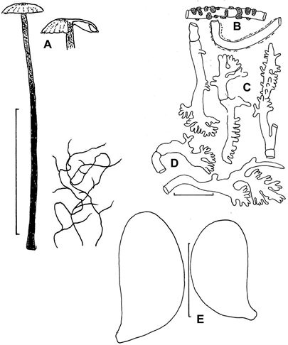
Fig. 36. Pseudomarasmius pallidocephalus (TENN-F-063098). A. Basidioma and rhizomorphs.
B–D. Pileipellis elements. B. Encrusted hyphal segment and hypha with slime sheath. C. Diverticulate hyphae. D. Diverticulate hyphal termini. E. Basidiospores. Scale bars: A = 20 mm. B–D. = 10 µm. E = 5 µm.
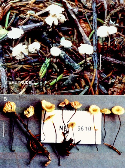
Fig. 37. Pseudomarasmius pallidocephalus. Basidiomata. A. Photo from nature (TENN-F-066344).
Courtesy Steve Trudell. B. Field-dried basidiomata (TENN-F-052401). Scale bars: 10 mm.
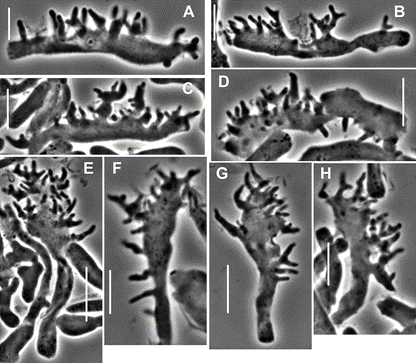
Fig. 38. Pseudomarasmius pallidocephalus (TENN-F-020486). Pileipellis elements. A–D. Repent, diverticulate hyphae. E–H. Broom cell-like, diverticulate hyphal termini. Scale bars: 10 µm.

Fig. 39. Pseudomarasmius pallidocephalus (MICH 51323). Pileipellis elements. A,B. Repent diverticulate hyphae. C–H. Broom cell-like, diverticulate hyphal termini. Note frequency of di- or trichotomously branched diverticula. Scale bars: 10 µm.

Fig. 40. Pseudomarasmius pallidocephalus (WTU-F-8911). Free-form pileipellis elements. A–C. Non-diverticulate individuals. D–I. Diverticulate individuals. Scale bars: 10 µm.
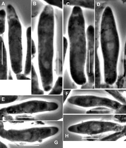
Fig. 41. Pseudomarasmius pallidocephalus.Pleurocystidia (A–D. MICH 51323. E–H. TENN-F-026266). Note frequent distal, vague partition of contents. Scale bars: 10 µm.
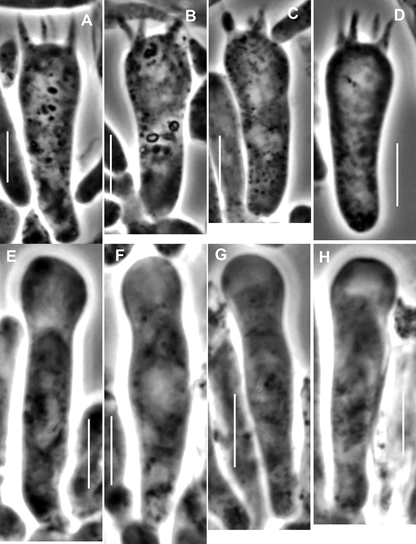
Fig. 42. Pseudomarasmius pallidocephalus. Hymenial structures. A–D. Basidia (MICH 51323). E–H. Cheilocystidia (TENN-F-026266).Scale bars: 10 µm.
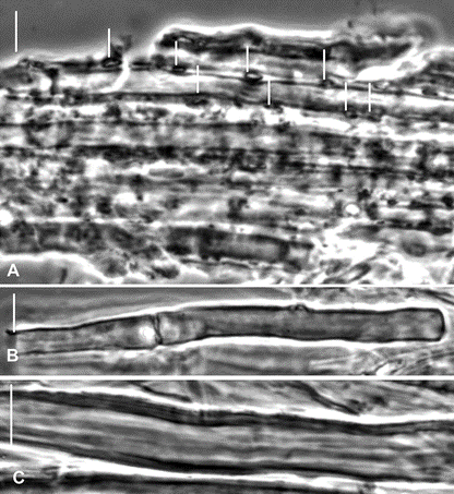
Fig. 43. Pseudomarasmius pallidocephalus (TENN-F-056761). Stipe medullary hyphae. A. Outer hyphae, with lines marking hyphal crust depositions. Stipe surface upward in photo. B, C. Individual inner medullary hyphae. B. Notes septum without clamp connection. C. Note wall thickness. Scale bars: 10 µm.
