Pseudofusicoccum adansoniae Pavlic, T.I. Burgess & M.J. Wingf., Mycologia 100(6): 855 (2008)
MycoBank number: MB 512048; Index Fungorum number: IF 512048; Facesoffungi number: FoF 00168; Fig. 1, 2
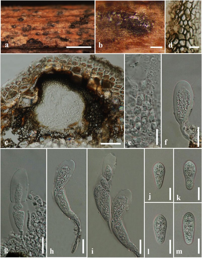
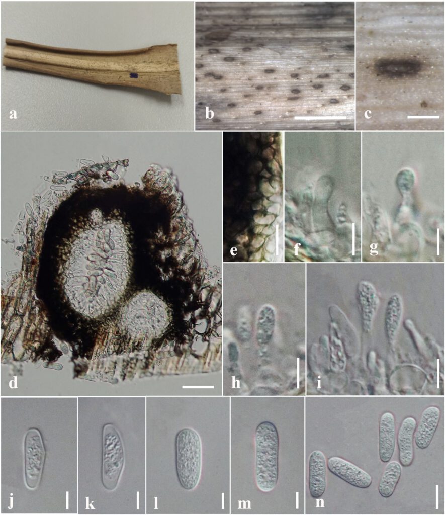
Saprobic on dead twig of Mangifera indica (sexual morph) and dead leaves of Epipremnum pinnatum (asexual morph). Sexual morph: Ascomata 160–170 μm high × 170–200 μm diam. (x̄ = 165 × 182 μm, n = 10), solitary, scattered, immersed, erumpent, globose to subglobose, gregarious, uniloculate. Peridium 20–50 μm wide, two-layered, outer layer composed of thick-walled, brown to dark brown cells of textura angularis, inner layer composed of thin-walled, hyaline cells of textura angularis. Hamathecium comprising 3–5 μm wide, hyaline, septate pseudoparaphyses, constricted at septa. Asci 50–140 × 20–25 μm (x̄ = 85 × 20 μm, n = 20), bitunicate, fissitunicate, 8-spored, cylindric-clavate to clavate, with a long pedicel, apically rounded with a well-developed ocular chamber. Ascospores 18–26 × 8–10 μm (x̄ = 22 × 9 μm, n = 30), 1–2-seriate, overlapping, hyaline, aseptate, short clavate, straight, smooth-walled, with granular appearance. Asexual morph: Coelomycetous. Conidiomata 175–200 μm high × 115–130 μm diam. (x̄ = 180 × 120 μm, n = 10), pycnidial, solitary, semi-immersed, uniloculate, subglobose to ellipsoid, black. Conidiomatal wall 30–38 μm wide, consist of 5–8 layers, outer layer composed of thick-walled, dark brown to brown cells of textura angularis, inner layer composed of thin-walled, hyaline to light brown cells of textura angularis. Conidiophores usually reduced to conidiogenous cells. Conidiogenous cells 5–6 × 2–4 μm (x̄ = 5.5 × 3 μm, n = 20), lining the pycnidial cavity, holoblastic, cylindrical, hyaline, discrete, determinate, smooth walled tapering to the apex. Conidia 18–21 x 5–8 (x̄ = 20 × 7 μm, L/W= 2.8, n = 20), aseptate, oblong, straight, occasionally slightly bent, hyaline, smooth-walled, with granular contents.
Material examined – Thailand, Chiang Rai Province, Mueang, Thasud, on a dead twig of Mangifera indica, 14 November 2020, Achala Rathnayaka, AN04 (MFLU 22-0181); ibid, Nang Lae village, on dead leaves of Epipremnum pinnatum, 13 May 2020, Achala Rathnayaka AV002 (MFLU 22-0182).
GenBank accession numbers – MFLU 22-0181: ITS: OP689652, LSU: OP689693; MFLU 22-0182: ITS: OP689653, β-tubulin: OP700654.
Known distribution (based on molecular data) – Australia (Pavlic et al. 2008), Thailand (Doilom et al. 2015, Senwanna et al. 2020, this study).
Known hosts (based on molecular data) – Adansonia gibbosa (Pavlic et al. 2008), Tectona grandis (Doilom et al. 2015), Hevea brasiliensis (Senwanna et al. 2020), Epipremnum pinnatum, Mangifera indica (this study).
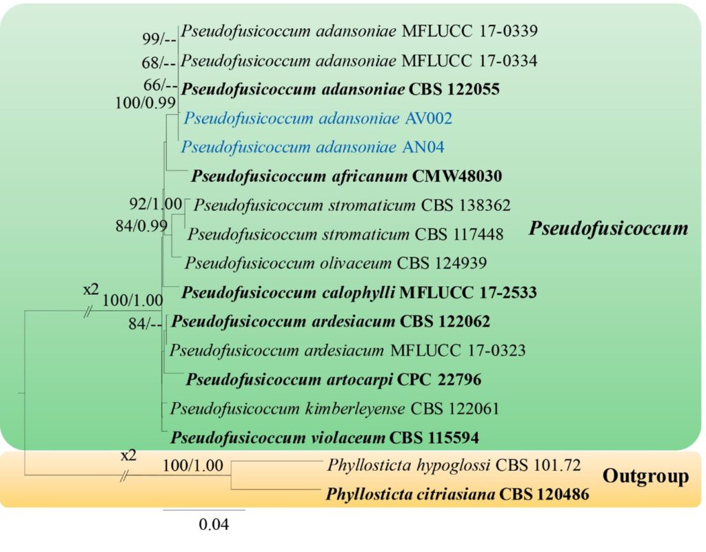
Notes – The morphologies of both sexual and asexual morphs of our fungal collections are similar to the holotype of P. adansoniae isolated from a canker on branches and twigs of Hevea brasiliensis (Senwanna et al. 2020) and dying branches of Adansonia gibbose in Australia (Pavlic et al. 2008). However, our sexual morph (MFLU 22-0181) has larger asci and ascospores (x̄ = 85 × 20 μm and x̄ = 22 × 9 μm) than those of the holotype (x̄ = 70 × 18 μm vs. x̄ = 16.6 × 6.3 μm) (Senwanna et al. 2020). Also, the length of conidia of our asexual morph (MFLU 22-0182) (x̄ = 20 × 7 μm, L/W= 2.8) is comparatively smaller than the holotype (x̄ = 22.5 × 5.2 μm, L/W= 4.3) (Pavlic et al. 2008). According to the phylogenetic analyses, our collections (MFLU 22-0181 and MFLU 22-0182) clustered with other P. adansoniae strains (CBS 122055, MFLUCC 17-0334, MFLUCC 17-0339) with 100% MPBS and 0.99 BYPP (Fig. 35).

Fig 1: Sexual morph of Pseudofusicoccum adansoniae (MFLU 22-0181, a new host record). a, b Appearance of ascostromata on host surface. c Section through ascomata. d Section through peridium. e Pseudoparaphyses. f–i Asci. j–m Ascospores. Scale bars: a = 1 mm, b = 100 μm, c = 50 μm, d, e, j–m = 10 μm, f–i = 20 μm.

Fig. 2. Asexual morph of Pseudofusicoccum adansoniae (MFLU 22-0182, a new host record). a Host. b, c Appearance of conidiomata on host tissue. d Vertical section of conidiomata. e Section through peridium. f−i Developing conidia attached to conidiogenous cells. j−n Conidia. Scale bars: b = 1 mm, c = 200 µm, d = 50 µm, e = 5 µm, f−i, n = 10 µm, j−m = 5 µm.

Fig. 3 — Phylogram generated from the maximum likelihood analysis based on the combined ITS, LSU, TEF1-α and β-tubulin sequence data of the genus Pseudofusicoccum. Seventeen strains are included in the combined analyses. Tree topology of the maximum likelihood analysis is similar to the Bayesian analysis. The best RAxML tree with a final likelihood value of -4983.29 is presented. Evolutionary models applied for all genes are GTR+I+G. The matrix had 285 distinct alignment patterns, with 32.63% of undetermined characters or gaps. Bootstrap support values for ML greater than 60% and Bayesian posterior probabilities greater than 0.90 are given near nodes, respectively. The tree was rooted with Phyllosticta citriasiana (CBS 120486) and P. hypoglossi (CBS 101.72). Ex-type strains are in bold. The newly generated sequences are indicated in yellow.
