Pseudodactylaria Crous, Persoonia 39: 421 (2017)
Index Fungorum number: IF 823411; MycoBank number: MB 823411; Facesoffungi number: FoF 05319; 2 species with sequence data.
Type species – Pseudodactylaria xanthorrhoeae Crous
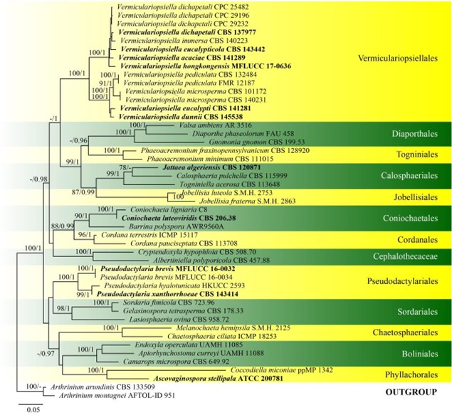
Notes – Pseudodactylaria was introduced by Crous et al. (2017a) for two dactylaria-like species, P. hyalotunicata and P. xanthorrhoeae, in the subclass Sordariomycetidae and class Sordariomycetes (Fig. 22). The genus is characterised by distinct, single, hyaline, unbranched and septate conidiophores, terminal, integrated, polyblastic, denticulate conidiogenous cells and solitary, fusoid-ellipsoid, hyaline conidia (Lin et al. 2018). Pseudodactylaria brevis is illustrated below.

Figure 22 – Phylogram generated from maximum likelihood analysis based on combined LSU, SSU, ITS and rpb2 sequence data of Pseudodactylariales and Vermiculariopsiellales. Forty-seven strains are included in the combined analyses which comprised 4320 characters (865 characters for LSU, 1634 characters for SSU, 662 characters for ITS, 1159 characters for rpb2) after alignment. Arthrinium arundinis (CBS 133509) and Arthrinium montagnei (AFTOL-ID 951) (Apiosporaceae, Xylariales) are used as outgroup taxa. Single gene analyses were carried out and the phylogenies were similar in topology and clade stability. Tree topology of the maximum likelihood analysis is similar to the Bayesian analysis. The best RaxML tree with a final likelihood value of – 29256.470401 is presented. Estimated base frequencies were as follows: A = 0.249068, C = 0.239305, G = 0.280423, T = 0.231203; substitution rates AC = 1.369489, AG = 2.705642, AT =
1.345736, CG = 1.341396, CT = 6.599852, GT = 1.000000; gamma distribution shape parameter a = 0.220775. Bootstrap support values for ML greater than 75% and Bayesian posterior probabilities greater than 0.95 are given near the nodes, respectively. Ex-type strains are in bold.

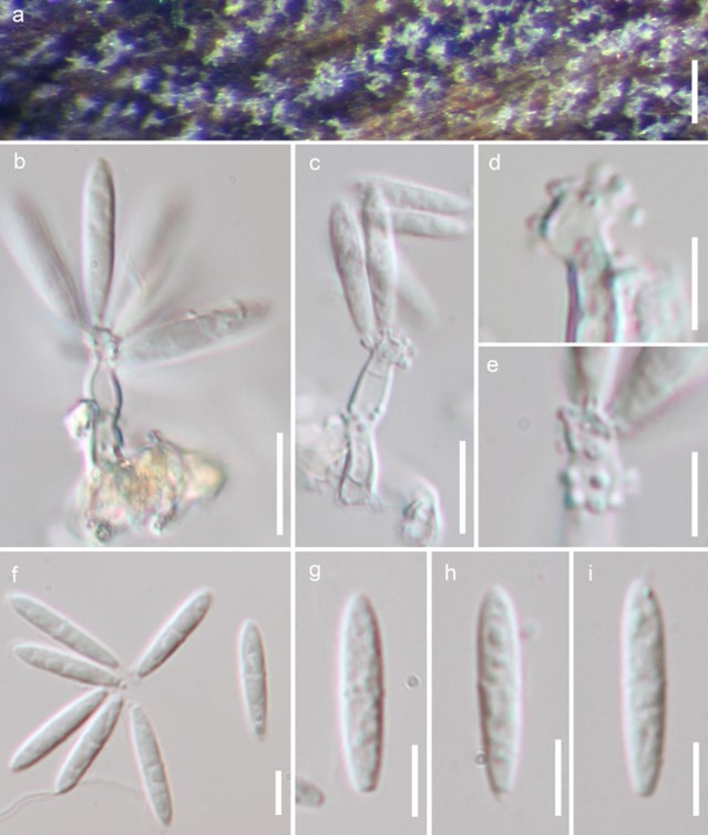
Figure 209 – Pseudodactylaria brevis (Material examined – THAILAND, Krabi, Wat Thum Sua, on decaying wood, 15 December 2015, S. Tibpromma M 1-5, MFLU 18-1038; HKAS 102202, paratype; living culture MFLUCC 16-0034). a Conidiophores on the host surface. b, c Conidiophores, conidiogenous cells with denticles and conidia. d, e Conidiogenous cells with denticles. f-i Conidia. Scale bars: b, c = 10 μm, d-i = 5 μm.
Table 1. Morphological comparisons among Pseudodactylaria species
|
Taxon |
Conidiophore size (µm) |
Conidiophore color |
Conidiogenous cells size (µm) |
Conidia size (µm) |
References |
| Pseudodactylaria albicolonia | 43–85 × 3–5.5 | Hyaline, sometimes light brown at the base | 18–51 × 3–6 | 14–19.5 × 2.5–4 | Boonmee et al. (2021) |
| P. aquatica | 40–100 × 3.5–4.5 | Brown to dark brown, paler towards the apex | 11.5–22 × 3.0–4.5 | 20–23 × 2.5–3.5 | Bao et al. (2021a, b) |
| P. brevis | Reduced to conidiogenous cells | Hyaline | 11–25 × 2–5 | 11.5–17.5 × 2.5–4.0 | Lin et al. (2018) |
| P. camporesiana | 35–45 × 3.5–5 | Brown, hyaline at upper part | 20–30 × 4–4.6 | 18–22 × 3.5–4.5 | Hyde et al. (2020b) |
| P. denticulata | 70–235 × 3–5.5 | Brown, paler towards the apex | 7–18 × 3–4 | 21–28 × 2–3.5 | This study |
| P. fusiformis | 30–50 × 3.5–4.5 | Hyaline | 11–25 × 2–5 | 11.5–17.5 × 2.5–4.0 | Lu et al. (2020) |
| P. hyalotunicata | 30–60 × 4–5 | Hyaline | – | 20–25 × 2.5–3 | Tusi et al. (1997) |
| P. longidenticulata | (55–)80–130(–175) × 3–5 | Brown, paler to hyaline towards the apex | 20–145 × 2.5–5 | 18–27 × 3–4.5 | This study |
| P. uniseptata | 90–185 × 3–5 | Dark brown, paler to hyaline towards the apex | 27–50 × 2.5–4 | 19–25 × 2.5–4 | This study |
| P. xanthorrhoeae | 20–50 × 4–5 | Hyaline | 5–30 × 3–4 | (20–)22–27(–33) × (3–)3.5(–4) | Crous et al. (2017) |
Species
