Pseudodactylaria denticulata J. Yang, E.B.G. Jones & K.D. Hyde, in Yang, Liu, Jones, Hyde, Liu, Bao, Liu, Li, Shen, Yu & Liu, Fungal Diversity 119: 164 (2023)
Index Fungorum number: IF 559822; MycoBank number: MB 559822; Facesoffungi number: FoF 12833
Etymology – referring to the denticulate conidiogenous cells
Holotype – MFLU 22-0068
Saprobic on decaying submerged wood in freshwater. Asexual morph: Colonies on wood effuse, hairy, scattered or aggregated, brown, with glistening conidial masses at the apex. Mycelium partly superficial, partly immersed, composed of septate, smooth, pale brown to hyaline hyphae. Conidiophores macronematous, mononematous, erect, straight or slightly flexuous, cylindrical, smooth-walled, septate, unbranched, brown, paler towards the apex, thick-walled, 70–235 × 3.2–5.6 µm (x̄ = 135 × 4 µm, n = 25). Conidiogenous cells polyblastic, integrated, terminal, cylindrical, pale brown to subhyaline, 7–18 µm long and 3.2–3.8 µm wide, denticulate, with up to 10 denticles at the apex. Conidia acrogenous, narrowly fusiform, uniseptate, smooth, guttulate, hyaline, 21–28 × 2.2–3.5 µm (x̄ = 24 × 3 µm, n = 25), thin-walled, sometimes with an inconspicuous appendage. Sexual morph: Undetermined.
Culture characteristics – Conidia germinating on PDA medium within 24 h. Germ tubes produced from both ends. Colonies on MEA medium slow growing, reaching 5–10 mm diam. after 2 months at 25 °C in natural light, with dense mycelium on the surface, grayish brown in the middle, white in the inner ring and brown in the outer ring; in reverse brown in the middle and paler at the entire margin.
Material examined – THAILAND, Trat Province, Amphoe Ko Chang, 12.133° N, 102.633° E, on decaying wood submerged in a freshwater stream, 27 April 2017, Y.Z. Lu, YJT18-2 (MFLU 22-0068, holotype; HKAS 112154, isotype), ex-type cultures MFLUCC 17-2125 and GZCC 20-0390.
Notes – Pseudodactylaria denticulata resembles P. aquatica, P. camporesiana, P. longidenticulata and P. uniseptata in having brown conidiophores, polyblastic denticulate conidiogenous cells and hyaline, narrowly fusiform, uniseptate conidia (Hyde et al. 2020b; Bao et al. 2021b; this study). The other species in the genus, P. albicolonia, P. brevis, P. fusiformis, P. hyalotunicata and P. xanthorrhoeae are distinct from the above species by hyaline conidiophores (Tusi et al. 1997; Crous et al. 2017; Lin et al. 2018; Lu et al. 2020; Boonmee et al. 2021). Pseudodactylaria denticulata is distinguished from P. aquatica, P. camporesiana, P. longidenticulata and P. uniseptata by longer conidiophores, shorter conidiogenous cells, and longer but slightly narrower conidia (Table 7). The phylogenetic analysis showed that Pseudodactylaria denticulata (MFLUCC 17-2125) clustered as a sister taxon to P. aquatica (MFLUCC 18-0201) with strong statistical support (100% ML BS/1.0 PP) (Fig. 53). Comparison of the ITS gene region revealed 20 bp (506/526 bp, six gaps) differences between P. denticulata and P. aquatica. We, therefore, recognize P. denticulata as a new species following the guidelines of Jeewon and Hyde (2016).
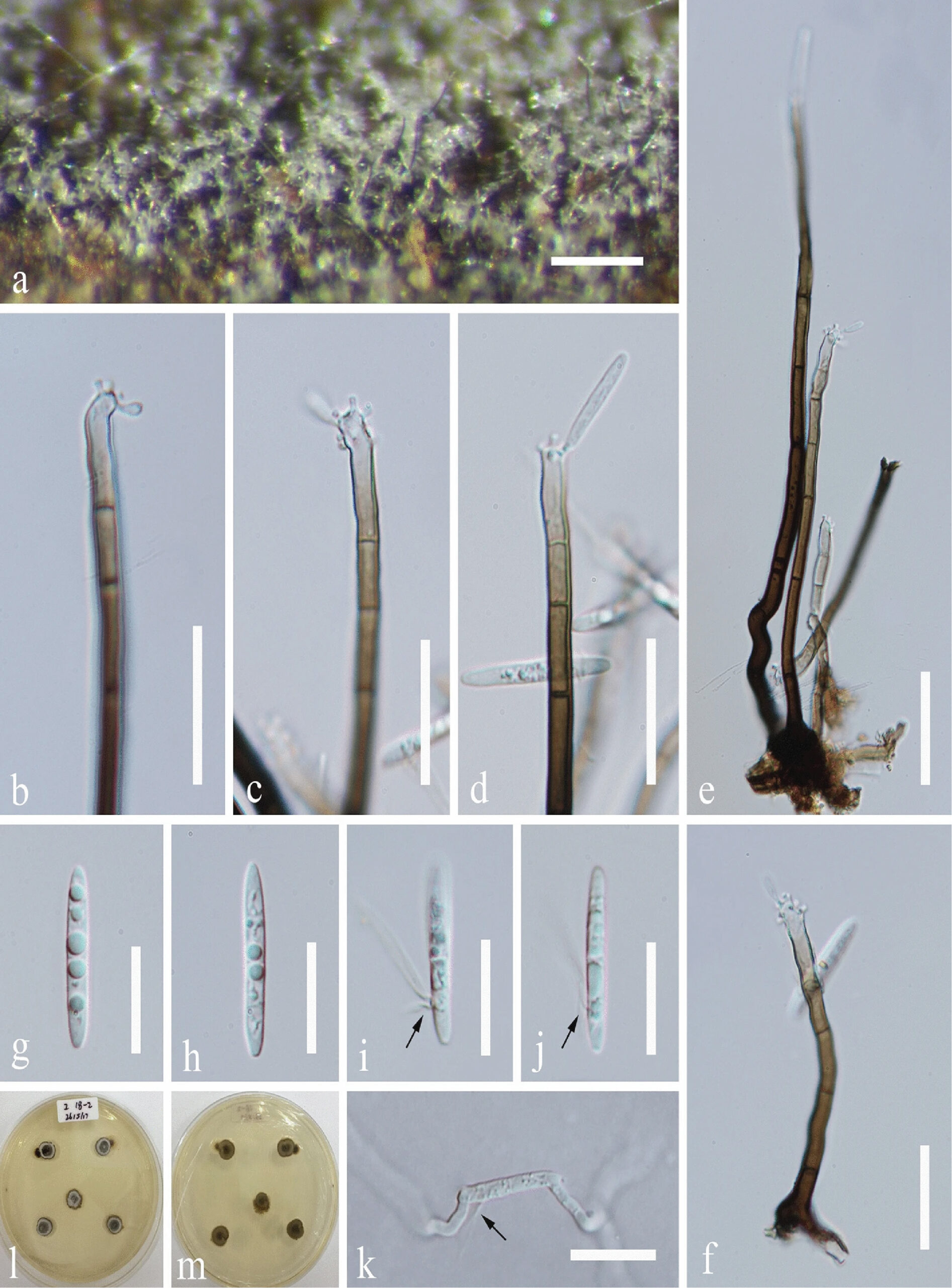
Figure 1. Pseudodactylaria denticulata (MFLU 22-0068, holotype) a Colony on wood. b–d Conidiogenous cells with conidia. e, f Conidiophores. g–j Conidia, arrows indicate appendages. l, m Culture, l from above, m from below. Scale bars: a = 200 µm, e = 30 µm, b–d, f, k = 20 µm, g–j = 15 µm