Pseudocercospora tamarindi Goon. & K.D. Hyde.
Index Fungorum number: IF550996, Facesoffungi number: FoF00473; Fig. 1
Etymology – in reference to the host Tamarindus indica.
Holotype – MFLU 14–0805
Pathogen on living leaves of Tamarindus indica. Sexual morph Undetermined. Asexual morph Leaf spots epiphyllous, indistinct, subcircular but mostly irregular, 0.5 – 1 cm diam., at most 2 cm diam. when confluent, pale brown to golden brown, delimited by leaf mid rib. Caespituli amphigenous but predominantly epiphyllous, abundant on leaf spots, dense, dark, punctiform. Stromata superficial, globular, pale to dark brown 11 – 25 μm diam., cells rounded, oblong or irregular, 2 – 3 μm diam., wall thickness less than 0.3 μm. Conidiophores fasciculate, arising from the stromata, brown becoming pale brown towards the apex, smooth, 0–2- septate, often short or one-cells, straight to slightly curved, apex rounded to truncate, unbranched, 3 – 12 × 1 – 2 μm (x̄ = 7.37 × 1.81 μm, n = 20), wall thickness less than 0.25 μm. Conidiogenous cells 5 – 7 × 0.8 – 2 μm (x̄ = 5.66 × 1.59, n = 10), integrated, terminal or conidiophores reduced to conidiogenous cells, occasionally branched, smooth, hyaline to pale brown, integrated, proliferating percurrently, conidiogenous loci inconspicuous. Conidia (13–) 17 – 25 (–30) × (1.8–) 2 – 3 (–3.5) μm (x̄ = 21 × 2.38 μm, n = 30), solitary, subhyaline to brown, cylindrical to obclavate, some showing constrictions at septa, some guttulate, subacute to obtuse apex, obconically subtruncate base, 2 – 14 – septate, hila neither thickened or darkened, 0.7 – 2 μm diam.
Culture characters – Colonies on PDA show slow growth, attaining 10 – 12 mm diam. after 3 weeks at 25 °C, dark-grey to black at the margin, grey to pale grey in the centre; reverse dark-green to black; medium dense with dense center, irregular, convex surface, smooth lobate margin, hairy, nonpigmented.
Material examined – THAILAND, Chiang Rai Province, Mueang Chiang Rai District, on living leaves of Tamarindus indica L. (Fabaceae), 12 February 2014, K.D. Hyde IG018 (MFLU 14–0805, holotype); ex-type living culture, MFLUCC 14–0805. GenBank ITS: KP744461; LSU: KP744506; SSU: KP753965.
Notes – The genus Pseudocercospora was introduced by Spegazzini (1910) with Pseudocercospora vitis as the type species. The genus comprises asexual morphs or species with Mycosphaerella-like sexual morphs, which includes many important plant pathogens (Hyde et al. 2013). They are characterized morphologically by their conidiophores and conidia being pigmented and by the conidiogenous loci being inconspicuous or at least unthickened and not darkened (Crous and Braun 2003; Crous et al. 2013). This new species is the first cercosporoid to be described from the host Tamarindus indica in Thailand. Previously, a species Cercospora tamarindi had been recorded on leaves of Tamarindus indica in Uttar Pradesh, India (Khan et al. 1988; Index Fungorum 2015). Pseudocercospora tamarindi differs from this species by having conidiophores that lack distinct spore scars with much less thick wall (>0.25μm) and darker coloured conidia. Although they both have a similar range for the number of septa (-14), the spores of P. tamarindi are much smaller (13–) 17 – 25 (–30) × (1.8–) 2 – 3 (–3.5) μm compared to those of C. tamarindi (38.4 – 93 × 3.8 μm according to Khan et al. 1988). A molecular analysis based on a combined gene analysis of ITS and LSU shows that Pseudocercospora tamarindi clusters within the major clade that includes the type species Pseudocercospora vitis with a bootstrap value of 84 % but is distant from the type species. Yet i t is clearly separated from other Pseudocercospora species. Pseudocercospora tamarindi is related with Pseudocercospora punctata (Wakef.) B. Sutton(= Cercostigmina punctata Wakef.) which has verruculose conidia (which the former lacks). It is also morphologically similar, but different in conidial size and the number of septa to Pseudocercospora ocimicola which has 1–8 septate conidia of (15–) 25 – 75 (–85) × 2 – 4 μm (Crous et al. 2013).
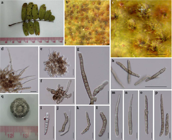
Fig. 1 Pseudocercospora tamarindi (holotype) a Symptoms on leaves (upper surface) b, c Close up of leaf spot d, e Stroma with attached conidiophores and conidia f Stromatal cells g-p Conidia of different sizes and maturity q Colony on PDA (3 weeks). Scale bars:b = 500 μm, c = 50 μm, d – f, h – o = 10 μm, g, p = 20 μm.