Pseudocamarosporium tiliicola Wijayaw., R.K. Schumach. & K.D. Hyde, in Wijayawardene et al., Cryptog. Mycol. 35(2): 193 (2014)
Index Fungorum Number: IF 550561; Facesoffungi number: FoF 000021
Etymology – Named after the host genus Tilia from which it was collected
Holotype – MFLU 14-0093
Saprobic in the bark of a dead and corticated branch of Tilia. Sexual state: Not observed. Asexual state: Conidiomata pycnidial, 670-700 µm diam., 650-720 µm high, solitary to gregarious, black, semi-immersed to superficial, unilocular, centrally papillate ostiole. Pycnidial wall multilayered, with 1-4 outer wall layers of dark brown cells of textura angularis, 5-10 cell layers in ostiole region, inner wall 2-4 layers, hyaline. Conidiophores reduced to conidiogenous cells. Conidiogenous cells with percurrent phialidic development, smooth, short, hyaline, segregated, formed from the inner layer of the pycnidial wall. Conidia 21-26 × 8-12 µm (› = 23.8 × 10.2 µm, n = 20), oblong to ellipsoid, straight to curved, rarely rounded, base tapered and seldom distinctly truncate, muriform, with 3-5 transverse septa and occasionally 2-3 longitudinal septa, brown to dark brown, smooth-walled.
Culture characteristics – on PDA white from above and pale brown from reverse, zonate, with thin mycelium, flat, circular, attaining a diam of 3 cm in 7 days at 18°C.
Material examined – Germany, near Berlin, on a branch of Tilia sp. (Malvaceae), 31 March 2013, Rene K. Schumacher, NNW G3 (G2/9) (MFLU 14-0093, holotype; isotype PDD 104442), living culture MFLUCC 13-0550 = ICMP 20372 = GUCC 0014.
Notes – Saccardo (1882) described Camarosporium tiliae Sacc. & Penz. from Tilia europaea L., however, Pseudocamarosporium tiliicola has larger conidia (21-26 × 8-12 µm vs 8-10 × 6-7 µm) and is therefore introduced as a new species.
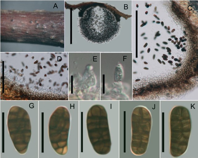
Fig. 9. Pseudocamarosporium tiliicola (holotype). A. Conidiomata on Tilia sp. B. Spore mass in cross section. C, D. Pycnidium wall. E, F. Different stages of developing conidia. G-K. Conidia. Scale bar: B. 650 µm; C. 300 µm; D. 120 µm; E, F. 15 µm; G-K. 20 µm