Pseudocamarosporium piceae Wijayawardene, E. Camporesi & K.D. Hyde, in Wijayawardene, Hyde, Bhat, Camporesi, Schumacher, Chethana, Wikee, Bahkali & Wang, Cryptog. Mycol. 35(2): 188 (2014)
Index Fungorum Number: IF 550559; Facesoffungi number: 000019
Etymology – Named after the host genus, Picea
Holotype – MFLU 14-0090
Saprobic on cones of Picea excels. Sexual state: Not observed. Asexual state: Conidiomata pycnidial, 120-140 µm diam., 110-130 µm high, mostly immersed, unilocular, solitary, scattered, moderately brown, dark brown at ostiolar papilla. Pycnidial wall multilayered, with 3-4 outer wall layers of dark brown cells of textura angularis, with inner layer with hyaline cells. Conidiophores reduced to conidiogenous cells. Conidiogenous cells formed from the inner layer of pycnidial wall, enteroblastic, with percurrent phialidic development, smooth, hyaline. Conidia 10-13 × 6-7 µm (› = 12.2 × 6.4 µm, n = 20), oblong, muriform broadly rounded at both ends, with 3 transverse and 1 longitudinal septa, smooth, variable in shape, hyaline when young, dark brown at maturity, smooth-walled.
Cultural characteristics – on PDA white to very light brown from above and reverse, with sparse mycelium, zonate, flat, circular, attaining a diam of 3.5 cm in 7 days at 18°C, reverse become dark green to black in 14 days.
Material examined – Italy, Forlì-Cesena Province, San Martino-Predappio, on dead cones of Picea excels (Pinaceae), 25 March 2012, E. Camporesi, NNW IT
308 (MFLU 14-0090, holotype; isotype PDD 104439), living culture MFLUCC 14-0192 = ICMP 20203 = GUCC 0012.
Notes – There are several coelomycetous taxa that have been recorded from Picea spp. (Sutton, 1980; Ellis & Ellis, 1985; Farr & Rossman, 2014), but the only Camarosporium species recorded is C. strobilinum E. Bommer et al. However, the conidial dimensions of these two taxa are distinct. Camarosporium strobilinum has smaller conidia (6 × 3-3.5 µm), while Pseudocamarosporium piceae has larger conidia (10-13 × 6-7 µm).
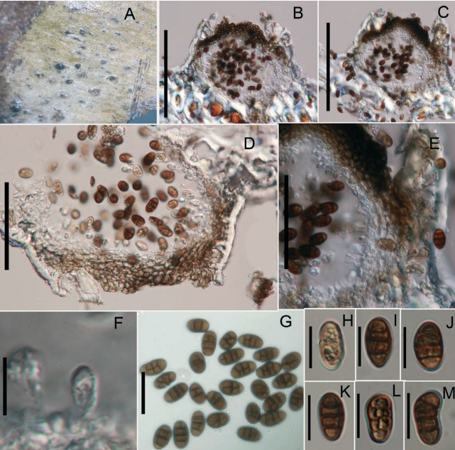
Fig. 1. Pseudocamarosporium piceae (holotype). A. Conidiomata on dead cone of Picea excels. B, C. Cross sections of pycnidium. D, E. Pycnidium wall. F. Developing conidia attach to conidiogenous cell. G-M. Conidia. Scale bar: B, C. 120 µm; D, E. 50 µm; F-M. 10 µm. Camarosporium-like species are polyphyletic in Pleosporales