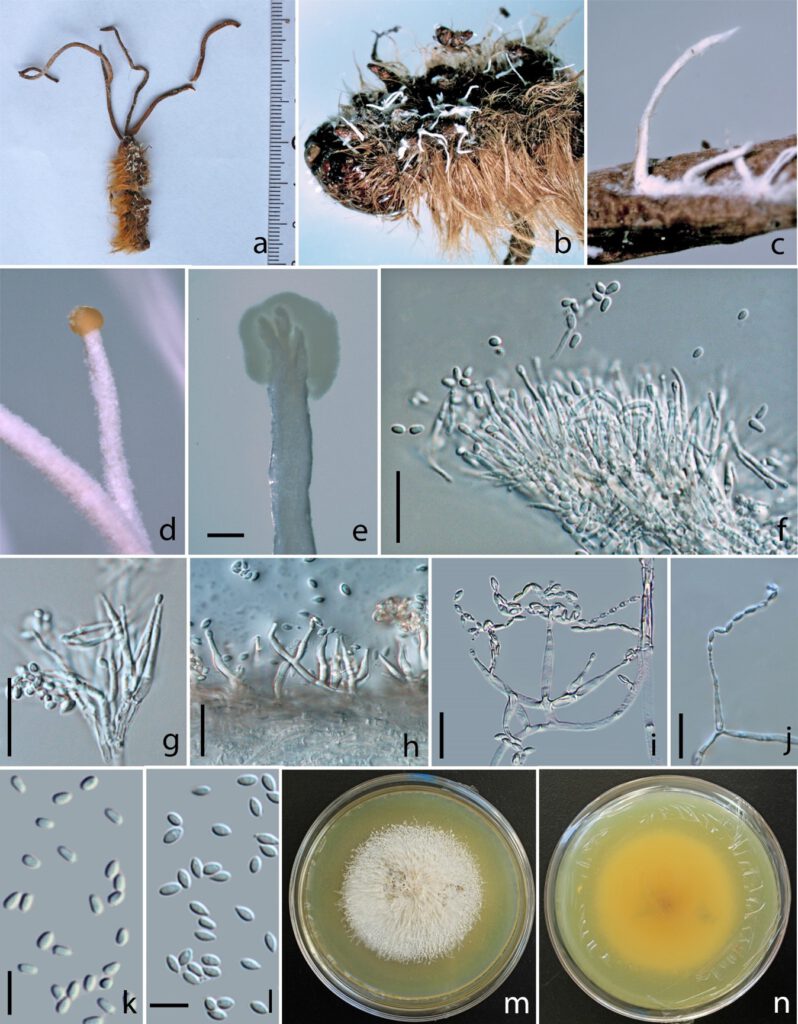
Pleurocordyceps heilongtanensis Y.B. Wang & Y.P. Xiao, sp. nov. Fig. 26.
MycoBank number: MB 559476; Index Fungorum number: IF 559476; Facesoffungi number: FoF 10742;
Etymology: The species name refers to the host species.
Holotype: kumcc3008
Hyperparasite on Ophiocordyceps sp. (Ophiocordycipitaceae), in the leaf litter. Asexual morph: Hyphomycetous. Synnemata 0.5–1 cm long, 1–2 mm wide, scattered on the surface of host, cylindrical, capitate, stipitate, unbranched, white, with or without fertile head. Stipe 0.5–1 cm long, 1–1.5 mm wide, cylindrical, white, with or without fertile head. Fertile head 1–2 mm wide, globose to subglobose, yellowish, with conidia masses. Conidiophores 14–26.4 μm ( = 20.2, n = 90) long, arranged in a parallel palisade-like layer on the surface of stipe or fertile head, hyaline, usually branched into 2–4 phialides. α-phialides 9.5–14.6 × 1.2–1.9 μm ( = 12.1 × 1.6 µm, n = 90), hyaline, smooth, elongated lageniform, caespitose, palisade-like, crowed, gathered in the top of synnema, mostly branched into2–4 phialides. β-phialides 8.4–12.5 × 1.2–2.1 μm ( = 10.5 × 1.7 µm, n = 90), hyaline, smooth, solitary, branched into 2 or 3 phialides, with or without metula at the base, directly growing from hyphae, , scattered along the stipe, lanceolate, ovate at the base, 4–9 μm long, tapering into a long neck, 2–5 μm long. α-conidia 2.1–3.1 × 1.5–2.3 μm μm ( = 2.6 × 1.9 µm, n = 90), one-celled, smooth-walled, subglobose to ovoid, occurring in the fertile head on the top of synnemata, yellowish slimy in mass. β-conidia 2.9–3.9 × 1.6–2.6 μm ( = 3.4 × 2.1 µm, n = 90), fusiform, along stipe of the synnema, one-celled, smooth-walled, hyaline.
Colonies on PDA medium, attaining 3 cm in 20 days at 25°C, white, with synnemata on the surface, reverse yellowish. Synnemata 2–10 mm long, 0.1–1 mm wide, emerging after 15 days in the margin of the colony, single, with or without fertile head. Fertile head 1–2 mm wide, globose to subglobose, yellowish, with conidia masses. Conidiophores 18–28.4 μm ( = 23.2, n = 90) long, arranged in a parallel palisade-like layer on the surface of stipe or fertile head, hyaline, usually branched into 2–4 phialides. α-phialides 10.5–18.6 × 1.4–1.9 μm ( = 14.6 × 1.7 µm, n = 90), hyaline, smooth, elongated lageniform, caespitose, palisade-like, crowed, gathered in the top of synnema, mostly branched into2–4 phialides. β-phialides 12.4–16.5 × 1.4–2.2 μm ( = 14.5 × 1.8 µm, n = 90), hyaline, smooth, branched into 2 or 3 phialides, with or without metula at the base, directly growing from hyphae, scattered along the stipe or on the surface of the colony, lanceolate, ovate at the base, 10–12 μm long, tapering into a long neck, 2–8 μm long. α-conidia 2.4–3.1 × 1.6–2.4 μm μm ( = 2.8 × 2 µm, n = 90), one-celled, smooth-walled, subglobose to ovoid, occurring in the fertile head on the top of synnemata, yellowish slimy in mass. β-conidia 3.4–4.8 × 1.7–2.6 μm ( = 4.1 × 2.2 µm, n = 90), fusiform, along stipe of the synnema, one-celled, smooth-walled, hyaline.
Material examined: China, Yunnan Province, Kunming City, Yeya Lake, parasitic on larvae of Lepidoptera, on the leaf litter, 30 September 2021, Yuanbing Wang (Holotype: kumcc3008), ex-type, XXXX.
Notes: Pleurocordyceps heilongtanensis is branching off the clade formed by Pleurocordyceps sinensis, Polycephalomyces ramosus and Polycephalomyces tomentosus in the phylogenetic tree with strong support value (Fig 16: 99% ML / 1.00 PP). More data is needed for Polycephalomyces ramosus and Polycephalomyces tomentosus. Pleurocordyceps heilongtanensis is distinct from Pleurocordyceps sinensis in producing smaller α-phialides and subglobose to ovoid β-conidia (Table 11). Thus, we introduce Polycephalomyces heilongtanensis is as a new species under Polycephalomyces and identify the host as Ophiocordyceps sp.

Fig. 26. Pleurocordyceps heilongtanensis (Holotype: kumcc3008) a Overview of Pleurocordyceps heilongtanensis. b Host with white synnemata. c–e Synnemata. f Conidiophores. g α-phialides. h–j β-phialides. k α-conidia. l β-phialides. m Upper side of colony. n Back side of colony. Scale Bars: e = 200 µm, f–j = 10 µm, k, l = 5 µm.
