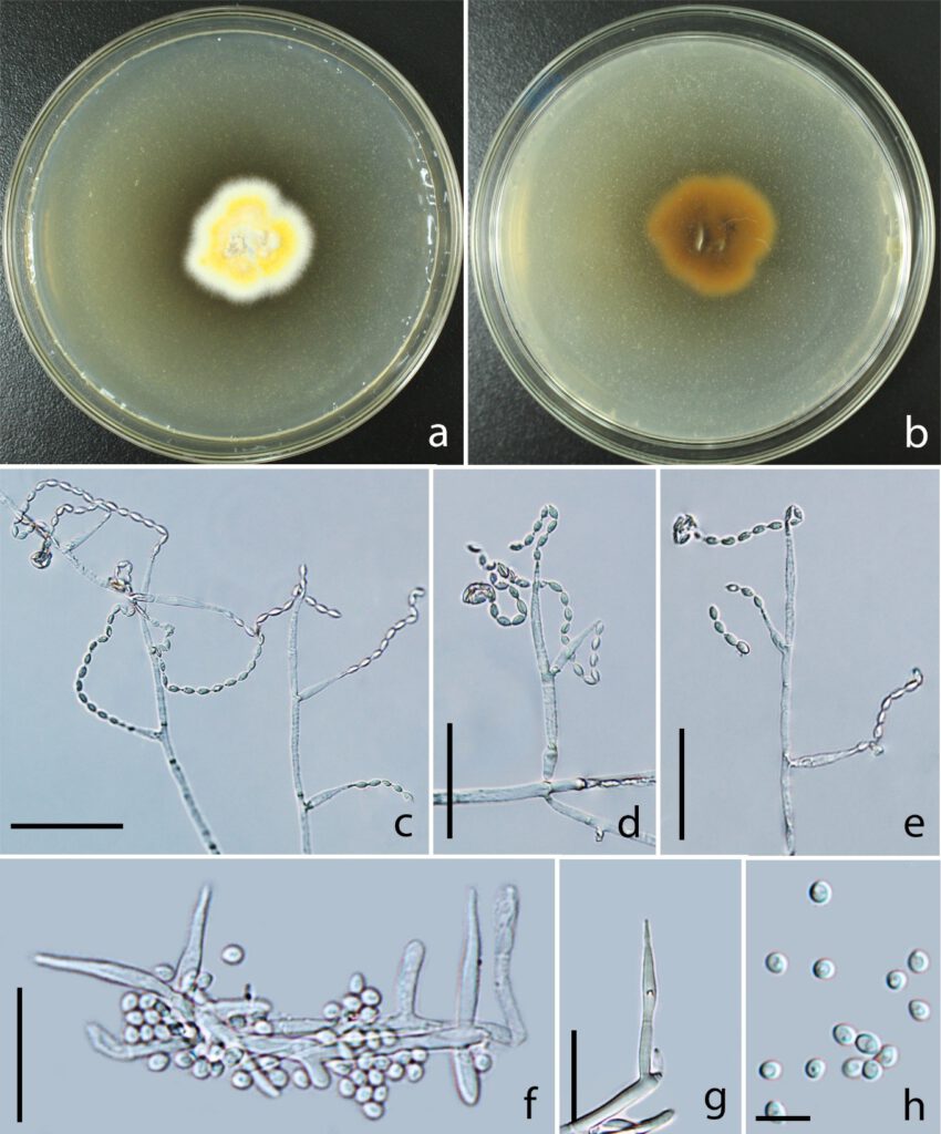
Pleurocordyceps vitellina Y.B. Wang & Y.P Xiao sp. nov. Fig. 27.
MycoBank number: MB 559477; Index Fungorum number: IF 559477; Facesoffungi number: FoF 10743;
Etymology: The species name refers to the egg yolk color of the colony.
Holotype: kumcc3005
Hyperparasite on Ophiocordyceps sinensis (Ophiocordycipitaceae), in the soil. Asexual morph: Hyphomycetous. Colonies on PDA medium, attaining 1.5 cm in 15 days at 25°C, egg yolk color in the middle, white on the edge, without synnemata on the surface, reverse brown. Conidial masses slime, light yellow, on the surface of colony. Conidiophores 32–42.4 × 1.3–2.2 μm ( = 37.2 × 1.8 µm, n = 90), hyaline, usually branched into 2 phialides, with or without metulae. Metulae 10–20.6 × 1.6–1.9 μm ( = 15.3 × 1.8 µm, n = 90), smooth, hyaline. Phialides two types. α-phialides 8.2–11.1 × 1.6–2.3 μm ( = 9.7 × 2 µm, n = 90), hyaline, smooth, elongated lageniform, crowed, gathered in the middle of colony. β-phialides 16.6–22.7 × 1.9–2.6 μm ( = 19.7 × 2.3 µm, n = 90), hyaline, smooth, directly growing from hyphae, with or without metula at the base, solitary, lanceolate, ovate at the base, 5–22 μm long, tapering into a short neck, 1–3 μm long. α-conidia 2.0–3.1 μm ( = 2.6 µm, n = 90) diam, one-celled, smooth-walled, spherical. β-conidia 3.8–5.1 × 1.8–2.4 μm ( = 4.5 × 2.1 µm, n = 90), fusiform, catenulate, one-celled, smooth-walled, hyaline.
Material examined: China, Yunnan Province, Kunming City, parasitic Ophiocordyceps sinensis (Ophiocordycipitaceae), in the soil, 30 September 2021, Yuanbing Wang, kumcc3008, (Holotype: kumcc3005), ex-type, kumcc3006, kumcc3007.
Notes: Pleurocordyceps vitellina branches off the clade formed by Pleurocordyceps agaricus and Polycephalomyces onorei (Fig. 16: 99% ML / 1.00 PP). Pleurocordyceps vitellina is distinct from other species of Pleurocordyceps in egg yolk color of colony, short α-phialides, large and catenulate β-conidia. Thus, we introduce Pleurocordyceps vitellina is as a new species under Pleurocordyceps and identify the host as Ophiocordyceps sinensis.

Fig. 27. Pleurocordyceps vitellina (Holotype: kumcc3005) a Upper side of colony. b Back side of colony. c Conidiophores. d, e β-phialides. f, g α-phialides. h α-conidia. Scale Bars: c–e = 20 µm, f, g = 10 µm, h = 5 µm.
