Plectosphaerella kunmingensis Phookamsak, J.F. Li & K.D. Hyde, sp. nov.; Index Fungorum number: IF556180
Etymology: The specific epithet ‘‘kunmingensis’’ refers to Kunming City, Yunnan, China where the species was
first collected.
Holotype: KUN-HKAS 102246
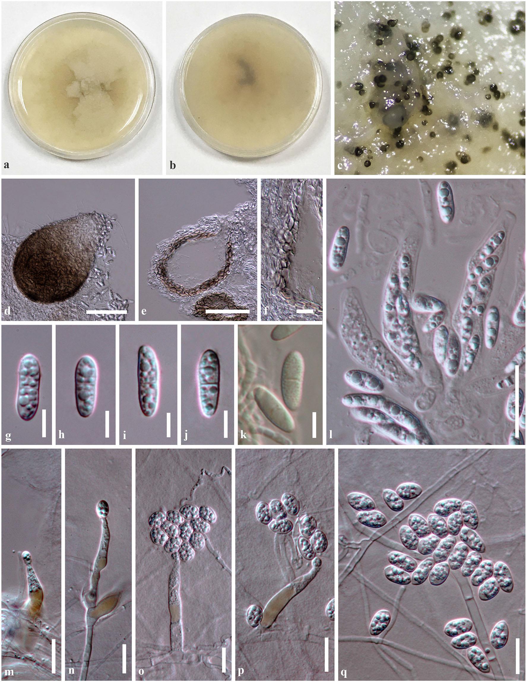
Colonies forming on PDA. Mycelium composed of 1–3 μm wide, septate, branched, smooth and thin-walled, hyaline hyphae, partly immersed on PDA. Sexual morph forming black, obpyriform ascomata after 8 weeks at 20–25 °C. Ascomata 100–185 μm high, 80–110 μm diam., perithecial, superficial or immersed in culture colonies, scattered, solitary to gregarious, subglobose to ovoid, or obpyriform, uni-loculate, glabrous, ostiole at centre, with a minute papilla. Peridium 7–18 μm wide, thin-walled, of equal thickness, composed of 1–3 layers, of dark brown pseudoparenchymatous cells, arranged in a textura angularis. Asci (42–)45–55(–62) × 10–13 μm (x̅ = 51.9 × 11.6 μm, n = 15), 8-spored, unitunicate, cylindric-obclavate to obpyriform, subsessile, thick-walled at apex, with J-, subapical ring. Ascospores (8–)10–13(–15) × 3–5 μm (x̅ = 11.9 × 4.3 μm, n = 50), overlapping 1–2-seriate, hyaline, ellipsoidal to subfusoid, with round ends, 1-septate, smooth-walled with small guttules, becoming finely echinulate when stained by Melzer’s reagent. Asexual morph: Hyphomycetous. Conidiophores 15–50(–58) × (1.5–)3–5 μm (x̅ = 38.5 × 4.2 μm, n = 20), macronematous or micronematous, produced from prostrate hyphae, lackinghyphal coils, sometimes branched, hyaline, yellowish brown at the base, thick-walled, smooth, septate, branched, straight or flexuous. Conidiogenous cells phialidic, determinate, integrated to discrete, hyaline, subcylindrical to ampulliform, smooth-walled, with periclinal wall thickening, with minute collarette. Conidia 7–10 × 4–7 μm (x̅ = 8.7 × 5.4 μm, n = 50), pleurogenous or acropleurogenous, formed in slimy heads at the apex of the phialides, hyaline, subglobose to ellipsoidal, aseptate, smooth, thin-walled, with granular contents. Chlamydospores not produced.
Culture characteristics: Colonies on PDA reaching 40–48 mm diam. after 3 weeks at 20–25 °C, dense, irregular in shape, flat to slightly raised, slightly rough at surface, with mycelia radiating outwards, sometimes forming sectors of different folds, fimbriate at edge, mucoid to floccose; from above, white yellowish to cream; from below, pale yellowish; not producing pigmentation in agar medium.
Material examined: CHINA, Yunnan Province, Kunming City, Kunming Institute of Botany, colonies forming on WA as a contaminated fungus, 5 July 2017, R. Phookamsak, KIB042 (dried culture herbarium: KUNHKAS 102246, holotype), ex-type living culture, KUMCC 18-0181. GenBank numbers: ITS = MH254296, LSU = MH254298, RPB2 = MH254297, TEF1-a = MH254295.
Notes: Phylogenetic analyses of a combined ITS, LSU and TEF1-a sequence dataset (Fig. 88) show the new species, Plectosphaerella kunmingensis forming a distinct lineage at the basal clade of Plectosphaerella in Plectosphaerellaceae with moderate support (76% ML and 0.96 BYPP). Plectosphaerella kunmingensis can be distinguished from other Plectosphaerella species in the lack of hyphal coils, as well as having partly coloured conidiophores. The sexual morph of P. kunmingensis differs from P. cucumerina in having smaller asci (P. kunmingensis, (42–)45–55(–62) × 10–13 μm versus 50–80 × 6–9 μm, P. cucumerina; Carlucci et al. 2012) and wider ascospores (P. kunmingensis, (8–)10–13(–15) 9 3–5 μm versus (9–)10.5–14(–15) × 2.5–3(–4) μm, P. cucumerina; Carlucci et al. 2012). Another species, Plectosphaerella sinensis was also found from China isolated from a healthy stem of Cucumis melo L. (Cucurbitaceae) (Su et al. 2017). Whereas, our isolate was found as a contaminated fungus on WA. Plectosphaerella kunmingensis has subglobose to ellipsoidal, aseptate conidia and lacks chlamydospores, while P. sinensis has ellipsoidal, 0–1-septate conidia with a slightly apiculate base, and forming intercalary or terminal, irregular, thick-walled chlamydospores (Su et al. 2017). In the BLASTn search on NCBI GenBank, the closest matches of ITS sequence of P. kunmingensis are P. nepalense (CBS 971.72) and P. cucumerina (XSD-75) with 95% similarities for both strains. In addition, a comparison of ITS nucleotide bases between P. kunmingensis and P. niemeijerarum (CBS 143233) shows that they differ in 42 base positions (8.08%/520 bp). Based on a molecular analyses coupled with morphological characteristics, we therefore, propose P. kunmingensis as a new species.
Fig. Plectosphaerella kunmingensis (KUN-HKAS 102246, holotype). a, b Culture characteristics on PDA (a = from above, b = from below). c Ascomata forming on PDA after 2 months. d Squash mount of the ascoma. e Section through ascoma. e, f Section through peridium. g–j Ascospores. k Ascospores stained in Melzer’s reagent. l Asci. m–p Conidiophores attached with conidia. q Conidia. Scale bars d, e = 50 μm, l = 20 μm, f, m–p, q = 10 μm, g–k = 5 μm.