Phylloporus gajari Hosen & Zhu L. Yang, Mycoscience 56: 585 (2015)
Index Fungorum number: IF803941
Basidiomata solitary or rarely caespitose, small to medium-sized. Pileus 21–47 mm in diam., plano-convex when young, becoming applanate in age, slightly depressed to depressed at center; surface dry, smooth when young, then becoming rough, finely areolate with age, mostly fox to English red (8D7–8), Sahara to caramel (6C5–6) around the edge when young, becoming yellowish brown or golden brown (5D7–8) to chamois (4C5–6) with age; margin straight to deflexed, usually uplifted when mature; context 4–8 mm thick at center, light yellow (1A4), slightly bluing when injured or cut. Hymenophore lamellate, deeply decurrent. Lamellae 15–26 × 2–8 mm, strongly intervenose to anastomosing, ventricose, subdistant, flexible; surface strongly transvenose, smooth, yellow (2A7) to pastel yellow (3A4), turning blue when injured; lamellulae common, in 1–2 tiers. Stipe 28–33 × 2–5 mm, central, straight to slightly curved, cylindrical; surface dry, fine yellow squamulose, fox to English red (8D7–8) when young, brownish orange (5C5–6) to brown (6E8); context solid, light yellow (1A4), slightly bluing then reddening with time in the upper part when cut or injured, but usually unchanging in the lower part. Basal mycelium well developed, tomentose, white. Odor and taste not distinctive.
Basidiospores [250/5/5] (9.2–)9.4–11.6–14.5(–14.9) × (4.4–)4.6–5.1–5.6(–5.8) µm, (1.76–)1.88–2.25–2.63(–2.9) , ellipsoid to fusoid or subfusoid, light yellow (1A4) to greyish yellow (1B4) when observed in H2O and 5% KOH, smooth under light microscope. Basidia 26–39 × 8–11 µm, 4-spored, with sterigmata 3–5 µm long, clavate, hyaline and light yellow (1A4) in H2O and 5% KOH. Hymenophoral trama 100–140 µm wide, bilateral, composed of hyaline hyphae 5–10 µm wide, cylindrical. Cheilo- and pleurocystidia 42–72 × 10–17 µm, narrowly clavate to clavate, hyaline to light yellow (1A4) in H2O and 5% KOH. Pileipellis a slightly interwoven trichoderm, composed of short chains of 4–5 cells, terminal cells 28–52 × 12–22 µm, cylindrical to somewhat slightly
inflated, smooth, hyaline to yellowish white (1A2) in 5% H2O and KOH. Pileal trama made up of hyaline hyphae 6–12 diam. Stipitipellis an interrupted hymeniderm, hyphae 6–13 μm diam; caulocystidia 22–46 × 13–21 µm, mostly clavate, sometimes mucronate, smooth, hyaline. Stipe trama composed of hyphae 8–11 µm wide, hyaline. Clamp connections not seen in any tissue.
Culture characteristics – uncultured.
Material examined – Thailand. Chiang Rai Province, Tha Sai District, Baan Hua Doi, 23/07/16, B. Chuankid, BC065 (MFLU16-2270); Chiang Mai Province, Muang District, N18°46′-E98°54′, 15/10/2015, O. Raspé & S. Vadthanarat, OR1184 (CMU-SDBR); Ubon Ratchathani Province, Muang District, N15°18′-E104°48′, 03/08/2015, O. Raspé & S. Vadthanarat, OR1104 and OR1110 (CMU-SDBR); —, N15°19’-E104°48’, 03/08/2015, O. Raspé & S. Vadthanarat, OR1108 (CMU-SDBR).
GenBank numbers – ATP6: MT861154 (OR1104), MT861153 (BC065), MT861155 (OR1108), MT861157 (OR1184), MT861156 (OR1110); ITS: MT880254 (OR1104); TEF1-α: MT861168 (OR1104), MT861167 (BC065), MT861169 (OR1108), MT861170 (OR1110); MT861171 (OR1184); RPB2: MT861161 (BC065), MT861162 (OR1108), MT861163 (OR1184).
Known distribution (based on molecular data) – Bangladesh (Hosen et al. 2015), Thailand (this study).
Known habitat (based on molecular data) – Soil in forests with monodominant stands of Shorea robusta (Hosen et al. 2015), dry dipterocarp forests (this study).
Notes – Phylloporus gajari is one of the most frequently collected species in forests dominated by Shorea robusta in Bangladesh. The pileus surface is pale red or reddish brown to yellowish brown or golden brown. The hymenophore is quickly staining blue when injured. The stipe is cylindrical, pale red to brownish orange or brown with white basal mycelium. The ITS sequences of the Thai specimen (OR1104) showed 100% similarity to P. gajari from Bangladesh (Hosen et al. 2015), which together with the morphological evidence, indicates that the studied specimens from Thailand belong in P. gajari. In the multi-gene phylogeny, the sequences from the Thai specimens clustered together with BS = 100%, PP = 1.0 and were clearly distinct from other Phylloporus species included in the analyses.
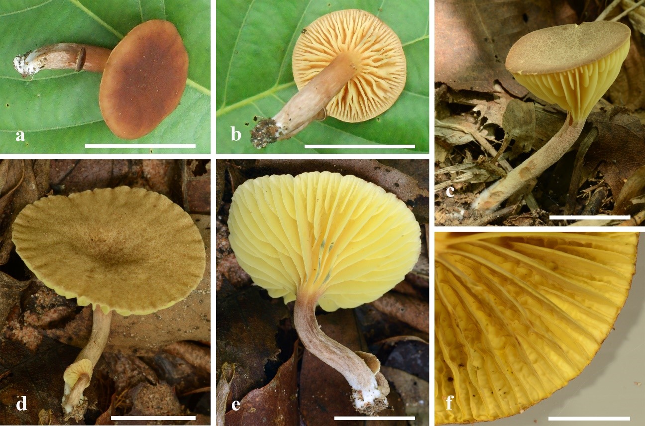
Fig. 1 – Basidiomata of Phylloporus gajari in the field. a, b Phylloporus gajari (BC065). c Phylloporus gajari (OR1184). d, e Phylloporus gajari (OR1108). f Close up of the hymenophore. Scale bars: a–e = 2 cm, f = 1 cm.

Fig. 2 – Microscopic characters of Phylloporus gajari (BC065). a Basidiospores. b Basidia. c Cheilo- and pleurocystidia. d Pileipellis. e Stipitipellis. Scale bars: a = 5 µm, b = 10 µm, c =20 µm, d–e = 50 µm.