Phlebia poroides Authority.
MycoBank number: MB 843327; Index Fungorum number: IF 843327; Facesoffungi number: FoF 12693;
Description
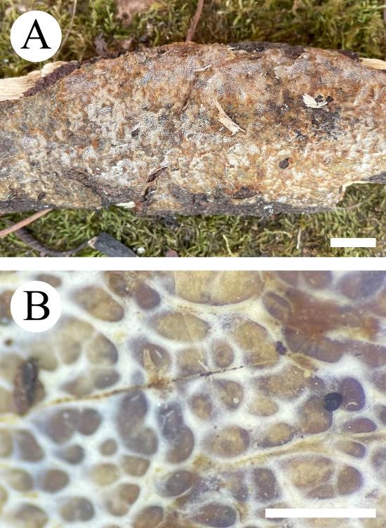
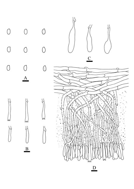
Basidiomata annual, resupinate, ceraceous, without odor or taste, when fresh, becoming hard and fragile upon drying, up to 11 cm long, 5 cm wide, 100–300 µm thick. Hymenophore poroid, buff when fresh, buff to slightly brown upon drying, 3–4/ mm, round, thin-walled, entire. Sterile margin narrow, slightly brown. Hyphal structure monomitic; generative hyphae clamped connections, colorless, thin-walled, IKI–, CB–; tissues unchanged in KOH. Subicular hyphae unbranched, 4–6.5 μm in diameter; subhymenial hyphae rarely branched, 2–4.5 μm in diameter; the presence of numerous brown sand-shaped substances among subhymenium. Hymenium cystidia pear-shaped, colorless, thin-walled, smooth, 21.9–47.3 × 7–10.5 μm, cystidioles absent; basidia cylindrical, with four sterigmata and a basal clamp connection, 16–28.6 × 3.2–4.9 μm. Basidiospores ellipsoid, colorless, thin-walled, smooth, often with 1–2 oil drops, IKI–, CB–, (3.2–)3.4–4.2(–4.5) × 1.7–2.5(–2.6) μm, L = 3.72 µm, W = 2.03 µm, Q = 1.77–1.83 (n = 60/2).
Material examined: China, Yunnan Province, Wenshan, Pingba Town, Wenshan National Nature Reserve, E 104°31′, N 23°22′, alt. 1720 m, on the fallen branch of angiosperm, 25 Jul 2019, C.L. Zhao, CLZhao 16121 (SWFC).
Distribution: The species is known from Yunnan Province, China, in a subtropical evergreen broad-leaved forest. It grows on moderately decayed angiosperm wood and causes white rot.
Sequence data: ITS: MW732405 (ITS5/ITS4); LSU: MW724797 (LROR/LR7); GAPDH: ON892533 (GAPDH-F/GAPDH-R); RPB1: ON892516 (RPB1-Af/RPB1-Cr); RPB2: ON918560 (bRPB2-6F/bRPB2-7.1R)
Notes: Phlebia poroides is sister to P. acerina Peck with lower supports, but P. acerina differs in its orange to brown hymenophore, and thick-walled subicular generative hyphae and smaller basidiospores (4.7–5.2 × 2–2 μm, Sytsma 1993).

Fig. 1 Basidioma of Phlebia poroides (holotype). — Scale bars: A = 1 cm, B = 1 mm.

Fig. 2 Microscopic structures of Phlebia poroides (drawn from the holotype). A. Basidiospores; B. Basidia and basidioles; C. Cystidia; D. A section of hymenium. — Scale bars: A = 5 μm, B–D = 10 μm.
