Phacidium chinaum G.C. Ren & K.D. Hyde sp. nov.
MycoBank number: MB; Index Fungorum number: IF; Facesoffungi number: FoF 10836, Fig. 2
Etymology: The species epithet reflects the country where the species was collected.
Holotype: KUN-HKAS 112899
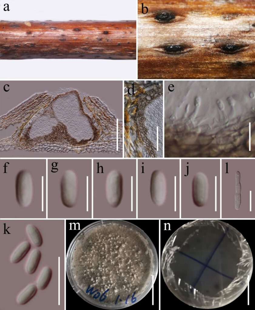
Saprobic on dead wood of Rosa sp. Sexual morph: Not observed. Asexual morph: Conidiomata 170–200 μm high, 150–240 μm diam. (x̅ = 180 × 200 μm, n= 5), black, pseudostromatic, solitary or gregarious, semi-immersed to superfcial, multi-locular, with 3–10 locules embedded in the pseudostroma. Ostioles 60–85 × 45–70 (x̅ = 73.5 × 58.5, n = 5) μm, centrally located, circular. Conidiomata wall 15–35 µm thick, 3–5 layered, comprising brown cells of textura angularis, thick-walled at basal, thin -walled at side. Conidiophores reduced to conidiogenous cells. Conidiogenous cells 3.5–5.5 × 1.2–2.0 µm (x̅ = 4.8 × 1.5, n = 10), hyaline, enteroblastic, phialidic, discrete, cylindrical, smooth-walled, arising from stratum. Conidia 4.5–6 × 2.0–2.4 µm (x̅ = 5.2 × 2.1 µm, n = 30), hyaline, oblong, unicellular, thick- and smooth-walled.
Culture Characters: Colonies on PDA, reaching 80–90 mm diam., after four weeks at 20–25 °C, medium dense, circular, rough, fluffy, cotton, gray, with white papillate on the surface, reverse dark-gray.
Material examined: China, Yunnan Province, Diqing Autonomous Prefecture, Xianggelila (27.28’8°N, 99.50’45°E), 2958m, on dead wood of Rosa sp. (Rosaceae), 30 August 2020. Guang-Cong Ren, W06 (KUN-HKAS 112899, holotype), ex-type living culture KUMCC 20-0168.
GenBank numbers: LSU: XXX, ITS: XXX, tef1-α: XXX, rpb2: XXX
Notes: Phacidium chinaum is introduced as a new species based on its distinct morphology, which is supported by phylogenetic analyses. In the phylogenetic analyses, P. chinaumi is distinct from extant species in Phacidium and formed a sister clade to Phacidium calderae, however, there is no bootstrap support (Figure 1). Phacidium chinaum is different to P. calderae in having oblong conidia and phialidic, cylindrical conidiogenous cells, while P. calderae in having subcylindrical conidia with apical mucoid appendage and proliferating with periclinal thickening conidiogenous cells (Crous et al. 2014).

Figure 2 Phacidium chinaum (KUN-HKAS 112899, holotype) a Herbarium specimen. b Conidiomata on the host. c Vertical sections of conidioma. d Sections of peridium. e conidiogenous cells and developing conidia. f–k Conidia. l Germinating conidium. m, n Culture on PDA. Scale bars: c = 100 µm. d = 50 µm. e, k = 10 µm. f–j = 5 µm. l = 20 µm. m, n= 30 mm.
