Pestalotiopsis umbrinospora Maharachch. & K.D. Hyde [as ‘umberspora‘], in Maharachchikumbura, Guo, Cai, Chukeatirote, Wu, Sun, Crous, Bhat, McKenzie, Bahkali & Hyde, Fungal Diversity 56(1): 121 (2012)
Index Fungorum number: IF 569202; Facesoffungi number: FoF 00016
Etymology – The specific epithet is based on the Latin = umber, in reference to the umber earth brown colour of the median cells of the conidia.
Conidiophores reduced to conidiogenous cells. Conidiogenous cells discrete or integrated, lageniform, hyaline, smooth-walled, and sometimes septate. Conidia 19–25 × 6–8 μm (x̄ = 21.3 × 6.5 μm), fusiform, straight to slightly curved, 4-septate; basal cell obconic to conic, hyaline or pale brown, thin and verruculose, 3–4.5 μm long (x̄ = 3.8 μm); three median cells 12–14 μm long (x̄ = 13.1 μm), umber brown to olivaceous, septa and periclinal walls darker than the rest of the cell, versicoloured, verruculose, second cell from base pale brown, 3–4.5 μm (x̄ =3.9 μm); third cell darker brown, 3.5–5 μm (x̄ = 4.3 μm); fourth cell darker, 3.5–4.5 μm (x̄ = 4.2 μm); apical cell 3–4.5 μm long (x̄ =3.9 μm), hyaline, conic to obconic; with apical appendages 22–35 μm long (x̄ = 27.7 μm), tubular, 1–3 (mainly 3), arising from the upper portion of the apical cell; basal appendage, 5–7 μm (x̄ = 5.9 μm), filiform.
Culture characters – Colonies on PDA reaching 7 cm diam. after 6 days at 25 °C, edge entire, whitish, aerial mycelium on the surface, fruiting bodies black, gregarious; reverse of culture pale yellow.
Habitat/Distribution – Saprobe on dead plant material, Guangxi Province, China.
Material examined – China, Guangxi Province, Shiwandashan, on dead leaves of unidentified plant, 30 December 1997, Wenping Wu WU1554j (HMAS042986, holotype; MFLU12-0421, isotype; ex-type living culture NN042986 = MFLUCC 12-0285).
Notes – Pestalotiopsis umberspora is a phylogenetically distinct species in the genus and separates well in tef1 and combined multi-locus tree with its phylogenetically related species P. crysea. Its umber-coloured and relatively wider mature conidia are characteristic of the species.
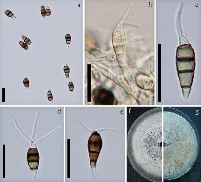
Figure 1. a. Pestalotiopsis umbrinospora (holotype). b. Conidiophores/ conidiogenous cells. c–e. Conidia. f. g. Colony on PDA, f. from above, g. from below. Scale Bars: a–e = 20 μm