Pestalotiopsis dracontomelon Maharachch. & K.D.Hyde, Fungal Diversity 72: 15 (2015)
Index Fungorum number: IF550943, MycoBank number: MB 550943; Facesoffungi number: FoF 00457;
Etymology – named after host genus, where it was isolated.
Holotype – MFLU 14–0207
Pathogen on Dracontomelon. Sexual morph Undetermined. Asexual morph Conidiomata pycnidial in culture on PDA, globose, aggregated or scattered, black, up to 150 μm diam. Conidiophores 2–3-septate, sparsely branched at the base, subcylindrical, up to 20μm long. Conidiogenous cells discrete or integrated, cylindrical, percurrently proliferating 1–3 times. Conidia 18–23 × 5.5– 7.5 μm (x̄ = 20 × 6.5 μm, n = 20), fusoid, ellipsoid, straight to slightly curved, 4-septate; basal cell conic with a truncate base, hyaline, rugose and thin-walled, 4–5 μm long; three median cells doliform, 13–17 μm long (x̄ = 15 μm), wall verruculose, concolourous, olivaceous, (second cell from the base 3.5–4.5 μm (x̄ = 4); third 3.5–4.5 μm (x̄ = 4 μm); fourth cell 3.5–4.5 μm (x̄ = 4 μm); apical cell 3–4 μm long, hyaline, subcylindrical, rugose and thin-walled; with 2–3 tubular apical appendages, arising from the apical crest, unbranched, filiform, flexuous 11–20 μm long (x̄ = 16 μm); basal appendage single, tubular, unbranched, centric, 2–7 μm long.
Culture characters – Colonies on PDA attaining 40–50 mm diam. after 7 d at 25 °C, with smooth edge, whitish, with sparse aerial mycelium on the surface with black, gregarious conidiomata; reverse similar in colour.
Material examined – Thailand, Chiang Rai, Nam Tak Huey Mesak Forest Park, on disease leaves of Dracontomelon dao Merr.&Rolfe (as D. mangiferum Blume), 10 February 2010, S.S.N Maharachchikumbura, SAJ-0011 (MFLU 14–0207, holotype); ex-type living culture, MFLUCC 10–0149.
GenBank Accession numbers – ITS: KP781877; TEF: KP781880.
Notes – Pestalotiopsis dracontomelon is a pathogenic species collected from leaves of Dracontomelon mangifera from Thailand. This species is a sister taxon to P. grevilleae and P. knightiae (Fig. 1). It differs from P. grevilleae and P. knightiae in having smaller conidia.
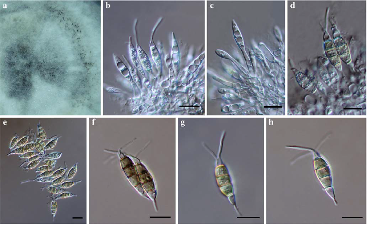
Fig. 1 Pestalotiopsis dracontomelon (holotype) a Conidiomata on PDA b–d Conidiogenous cells e–h Conidia. Scale bars: b-h=10μm.