Pestalotiopsis digitalis Maharachch & K.D. Hyde, Fungal Diversity 72: 14 (2015)
Index Fungorum number: IF550945, MycoBank number: MB 550945; Facesoffungi number: FoF 00459
Etymology – named after host genus, where it was isolated.
Holotype – MFLU 14–0208
Pathogen on Digitalis purpurea. Sexual morph Undetermined. Asexual morph Conidiomata pycnidial in culture on PDA, globose, scattered or gregarious and confluent, semi-immersed, dark brown, up to 100 μm diam. Conidiophores often reduced to conidiogenous cells. Conidiogenous cells discrete ampulliform to lageniform, smooth, thin-walled, hyaline, with 1–2 proliferations, sometimes remain vegetative. Conidia 18–22 × 7–9 μm (x̄ = 20 × 8.2 μm, n=20), fusiform, straight to slightly curved, 4-septate, basal cell conic to obconic, hyaline or slightly olivaceous, thin- and verruculose, 2–3.5 μm long (x̄ = 2.7 μm), with three median cells, doliform, concolourous, olivaceous, septa and periclinal walls darker than the rest of the cell, together 11– 17 μm long (x̄ = 15 μm) second cell from base 3–4.5 μm (x̄ = 4.1 μm); third cell 3–4.5 μm (x̄ = 4.1 μm); fourth cell 3– 4.5 μm (x̄ = 4.1 μm); apical cell hyaline, conic, 2–3.5 μm long (x̄ = 2.7 μm); with 1–3 tubular apical appendages (mainly 2), arising from the apex of the apical cell, 8–17 μm long (x̄ = 13 μm); basal appendage 4–7 μm long.
Culture characters – Colonies on PDA attaining 30–40 mm diam. after 7 d at 25 °C, with smooth edge, pale honey coloured, with dense aerial mycelium on the surface with black, gregarious conidiomata; reverse similar in colour.
Material examined – New Zealand, on leaf spots of Digitalis purpurea, 01 June 1972, J.M. Dingley 7270 (MFLU 14–0208, holotype); ex-type living cultures ICMP 5434.
GenBank Accession numbers – ITS: KP781879; TUB: KP781883.
Notes – Pestalotiopsis digitalis forms a sister clade to species including P. parva and P. rosea (Fig. 1). Pestalotiopsis rosea differs from P. dracontomelon in having distinctly narrow conidia. Furthermore, the reddish colony is unique to P. rosea and this reddish colour can be seen even in conidiogenous cells and some conidia. Furthermore, conidia of P. digitalis are longer than those of P. parva.
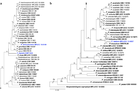
Fig. 1 Phylogram generated from Maximum Likelihood analysis based on combined ITS, β-tubulin and TEF gene regions of Pestalotiopsis. Maximum likelihood bootstrap support values greater than 50 % are indicated above or below the nodes. The ex-types (reference strains) are in bold; the new isolates are in blue. The tree is rooted with Neopestalotiopsis saprophyta MFLUCC 12-0282.