Paramicrosphaeropsis ellipsoidea L.W. Hou, L. Cai & Crous, sp. nov.
MycoBank number: MB 833539; Index Fungorum number: IF 833539; Facesoffungi number: FoF 11524; Fig. 42.
Etymology: Name refers to the ellipsoidal conidia of this species.
Description: Conidiomata pycnidial, semi-immersed, abundant, scattered or aggregated, mostly solitary, sometimes 2– 5 confluent, globose to subglobose, pale brown to dark brown, with hyphal outgrowths, (100 –)150 – 490 × (80 –)110– 440 μm.
Ostiole inconspicuous. Pycnidial wall pseudoparenchymatous, composed of isodiametric cells, thin, hyaline at beginning, dark brown with age, 4– 5 layers, 10– 30 μm thick, outer 1– 2 cell layers slightly pigmented. Conidiogenous cells phialidic, hyaline, smooth, subglobose, ampulliform or doliiform, 7– 11 × 5.5 – 10 μm. Conidia variable in shape, mostly broad ellipsoidal, some oblong or ovoid, straight or slightly curved, smooth- and thin-walled, hyaline, later becoming brown with age, aseptate, 5– 11 × 3.5 – 6.5 μm, with numerous minute guttules. Conidial matrix brownish.
Culture characteristics: Colonies on OA reaching 10 – 15 mm diam after 7 d at 25 °C, margin irregular, aerial mycelium flat, oliva- ceous, some zone formed pycnidia without aerial mycelium and in buff colour; reverse dark olivaceous with buff edge. Colonies on MEA 10 – 15 mm diam after 7 d, margin regular, aerial mycelium flat to floccose, pink to white, with some iron grey zone; reverse pale brown with buff edge, with some iron grey zone. Colonies on PDA reaching 10 – 15 mm diam after 7 d, margin regular, covered by flat aerial mycelium, rosy buff to pink, some brown pycnidia visible near the centre; reverse concolourous. NaOH spot test: a pale red-brown discolouration on OA.
Typus: Spain, from Quercus ilex (Fagaceae), 1996, R.F. Cas- ta~neda (holotype CBS H-23671, ex-type living culture CBS 194.97).
Additional material examined: Spain, from decayed twig of Quercus ilex (Fagaceae), 20 Jul. 1996, R.F. Casta~neda, culture CBS 197.97.
Notes: Isolates CBS 194.97 and CBS 197.97 were previously identified as “Microsphaeropsis sp.”, and characterised by producing brown conidia. However, based on the phylogenetic analysis (Fig. 1), these two isolates formed a distinct clade basal to Neomicrosphaeropsis and Microsphaeropsis. Therefore, we accommodated CBS 197.97 and CBS 194.97 as a new species in a novel genus, Paramicrosphaeropsis. Morphologically, Paramicrosphaeropsis differs from Neomicrosphaeropsis in its sub- globose, ampulliform or doliiform conidiogenous cells, while Neomicrosphaeropsis produces subcylindrical conidiogenous cells.
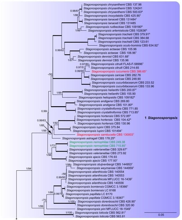
Fig. 1. Phylogenetic tree inferred from a Maximum Likelihood analysis based on a concatenated alignment of LSU, ITS, rpb2 and tub2 sequences of 597 strains representing Didymellaceae and outgroup sequences. The RAxML bootstrap support values (MLBS) above 50 % and Bayesian posterior probabilities (BPP) above 0.80 are given at the nodes (BPP/MLBS). Some of the basal branches were shortened to facilitate layout (the fraction in round parentheses refers to the presented length compared to the actual length of the branch). The scale bar represents the expected number of changes per site. Genera are delimited in coloured boxes, with the genus name indicated to the right. Strains with special status are indicated with a superscript letter after the accession number (R: representative; T: ex-type). The new species are printed in red font and new combinations in green font. The tree is rooted to Coniothyrium palmarum culture CBS 400.71, Neocucurbitaria aquatica culture CBS 297.74 and Pleiochaeta setosa cultures CBS 496.63 and CBS 118.25.
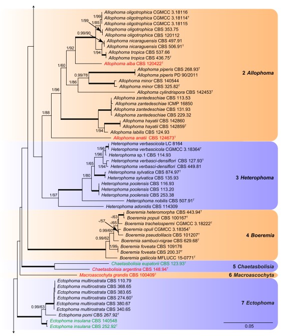
Fig. 1. (Continued).
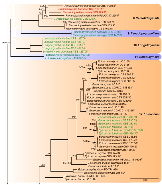
Fig. 1. (Continued).
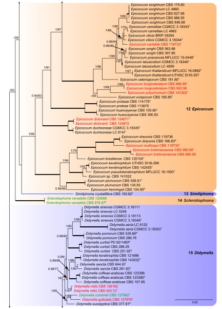
Fig. 1. (Continued).
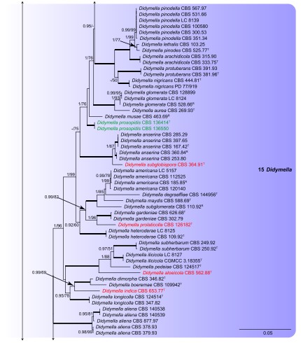
Fig. 1. (Continued).
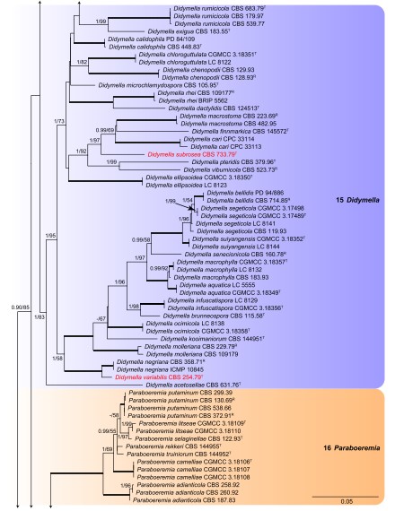
Fig. 1. (Continued).
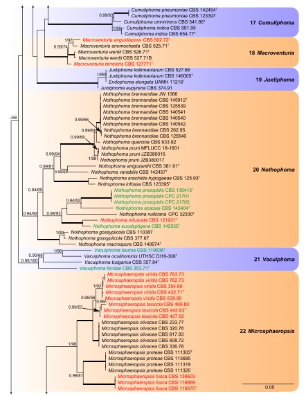
Fig. 1. (Continued).
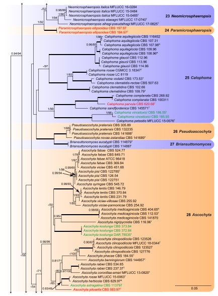
Fig. 1. (Continued).
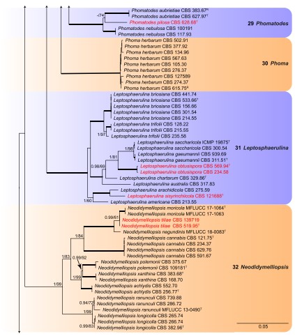
Fig. 1. (Continued).
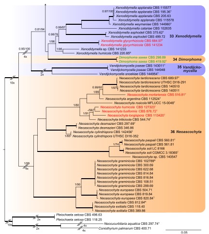
Fig. 1. (Continued).
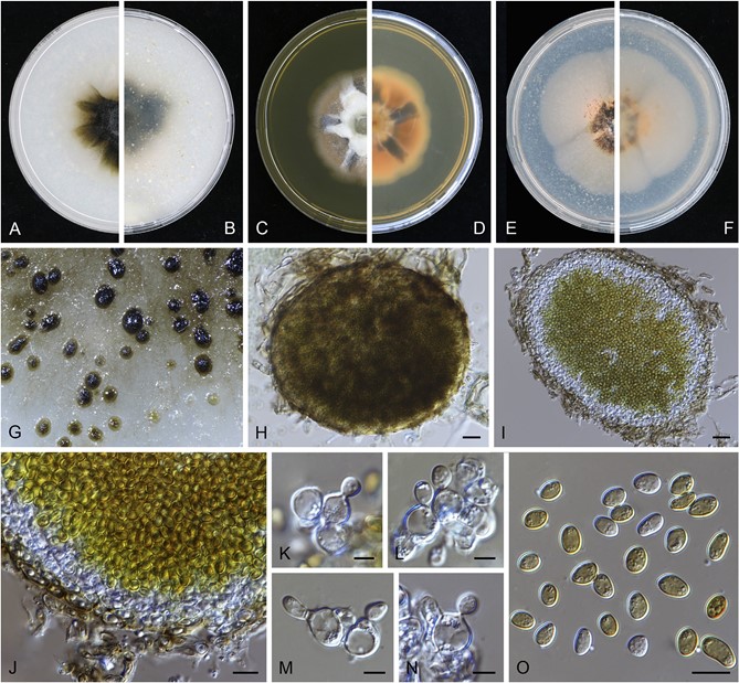
Fig. 42. Paramicrosphaeropsis ellipsoidea (CBS 194.97). A–B. Colony on OA (front and reverse). C–D. Colony on MEA (front and reverse). E–F. Colony on PDA (front and reverse). G. Pycnidia forming on OA. H. Pycnidium. I. Section through pycnidium. J. Section of pycnidial wall. K– N. Conidiogenous cells. O. Conidia. Scale bars: H–I= 20 μm; J, O = 10 μm; K– N = 5 μm.
