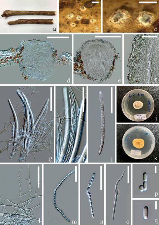
Ostropomyces pruinosellus Thiyagaraja, Lücking, Ertz and K.D. Hyde, in Thiyagaraja, Lücking, Ertz, Karunarathna, Wanasinghe, Lumyong and Hyde, Journal of Fungi 7(no. 105): 12 (2021) Figure 4.
MycoBank number: MB 556556; Index Fungorum number: IF 556556; Facesoffungi number: FoF 10097;
Saprobic on the dead stem of an unidentified climbing plant. Sexual morph: Ascomata 230–300 × 300–365 µm (x = 265 × 330, n = 5), perithecial, subglobose, unilocular, gregarious, immersed at first and raising the substrates into small pustules and later opening by entire pores, exposing the discs. Discs pale grey to olivaceous, convex and covered by thick pru- inose substance. Inner mass creamy-yellow. Exciple 25–50 µm (x = 40, n = 20), comprising interwoven and hyaline hyphae without crystalline inclusions and periphysoidal layers. Hymenium consisted of paraphyses and asci, lying parallel to the substrates. Paraphyses 1–3 µm (x = 2, n = 30) wide, anastomosing, aseptate, branched and longer than asci. Asci 180–265 × 9–15 µm (x = 220 × 12, n = 20), cylindrical, unitunicate and thick-walled, with a thickened cap. Apical caps 5–6.5 × 3–4.5 µm (x = 5.5 × 3.7, n = 5), hemispheric and pierced by a pore. Ascospores hyaline, filiform, as long as asci and easily disarticulating into part-spores in mature asci. Part-spores 2.5–6 2–3 µm (x = 4 × 2.5, n = 40), subglobose to ellipsoidal and unicellular, with two oil droplets. Asexual morph: Undetermined.
Culture characters: Culture was established from germinating part-spores. The colony was slowly grown on PDA media, reaching 3 cm after incubation for two months in 28 ◦C. It was sterile, yellowish white, nearly circular, dense, umbonate, surface folded, cottony and reverse creamy-yellow.
Material examined: Thailand, Chiang Rai Province, Doilan District, on dead stem of an unidentified dicotyledonous climbing plant, 27 March 2019, Deping Wei, CR2706 (MFLU 21-0115); living culture MFLUCC 21-0112.
Notes: Our isolate (MFLU 21-0115) resembles Ostropomyces pruinosellus (MFLU 20-0539) in having subglobose, white-pruinose, perithecioid ascomata, cylindrical asci and filamentous, anastomosing paraphyses and filiform ascospores that disarticulate into numerous ellipsoidal part-spores. Phylogenetic analyses supported close affinity of our isolates (MFLU 21-0115 and MFLUCC 21-0112) to Ostropomyces pruinosellus (MFLU 20- 0538), and the three were grouped together with maximum statistical support. Here, we consider our isolate as a new collection of Ostropomyces pruinosellus and provide additional molecular and morphological data as well as culture characteristics for this species.

Figure 4. Ostropomyces pruinosellus (MFLU 21-0115). (a) Substrate; (b,c) ascomata on host; (d,e) vertical sections through ascomata; (f) excipulum; (g–i) Asci; (j,k) upper and lower view of culture on PDA media after incubation for two months; (l) paraphyses; (m,n) catenulate ascospores;
(o) germinating ascospore; (p,q) part-spores. Scale bars: (b,c) = 500 µm; (d,e) = 200 µm; (g,i) = 100 µm; (h,l) = 50 µm; (f,m–o) = 30 µm; (p,q) = 5 µm.
