Novopuccinia Y. M. Liang & Y. Liu, gen. nov.
MycoBank number: MB 838356; Index Fungorum number: IF 838356; Facesoffungi number: FoF;
Type species: Novopuccinia sycopsis-sinensis Y. M. Liang & Y. Liu, sp. nov.
Etymology: Novopuccinia (Lat.) referring to the new type taxon in Puccinia-complex.
Descriptions: Spermogonia subepidermal and mostly globose. Aecia subepidermal in origin, cupulate with well-developed peridium (Aecidium-type). Uredinia subepidermal in origin and becoming erumpent (Uredo-type). Telia subepidermal in origin, becoming erumpent, either pulvinate or columnar; teliospores one-celled and catenulate with a long intercalary cell or two- celled and pedicellate.
Habitat and distribution: Currently only known on Hamamelidaceae in Asia.
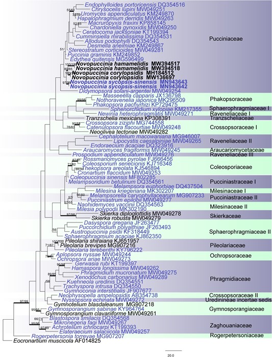
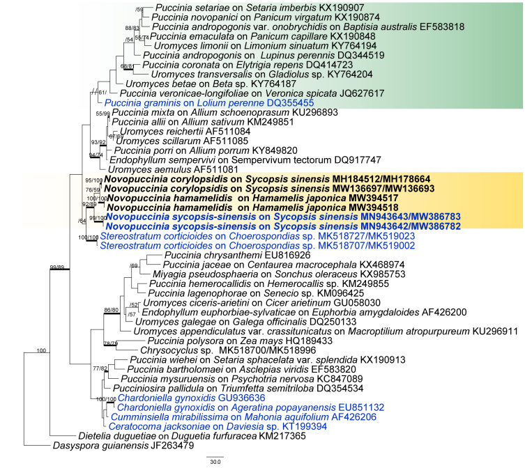
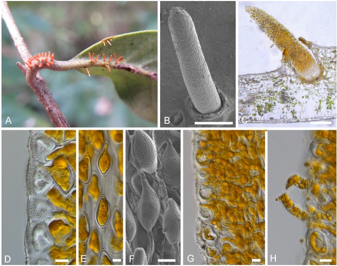
Notes: – Morphologically, the telia/teliospores of Novopuccinia consist of two types. One is represented by N. sycopsis-sinensis, which produces one-celled, catenulate teliospores in columnar telia. It is taxonomically distinct from other morphologically similar genera in Pucciniaceae, i.e., Ceratocoma, Chardoniella, Dietelia, and Trichopsora. Novopuccinia (Figure 4), Chardoniella, and Trichopsora produce hair-like telial columns and long, pedicel-like intercalary cells between individual teliospores (Lagerheim, 1891; Kern, 1939). In contrast, Ceratocoma and Dietelia have small, often inconspicuous, intercalary cells (Buriticá and Hennen, 1980; Buriticá, 1991; Cummins and Hiratsuka, 2003). The presence of a well-developed peridium in the telia is also a distinct characteristic of Novopuccinia, separating it from Ceratocoma, Chardoniella, and Trichopsora, which produce non-peridiate telia (Cummins and Hiratsuka, 2003; Berndt, 2018). A metabasidium replaces a probasidium without substantial morphological change in Trichopsora, while Ceratocoma, Chardoniella, and Novopuccinia produce a metabasidium external to a probasidium. In addition to these morphological distinctions, N. sycopsis-sinensis with one-celled teliospores is clearly separated from Ceratocoma, Chardoniella, and Dietelia in the molecular phylogenetic tree (Figures 2, 3: molecular data of Trichopsora for the analysis was lacking). Another telium/teliospore type is similar to those of Puccinia in two-celled with pedicellate (Figure 4), but it can be distinguished from almost all other species by thick-walled teliospores and Hamamelidaceae host. Our phylogenetic results also supported the independence of this type of rust on Hamamelidaceae from the large genus Puccinia (Figures 2, 3).

FIGURE 2 | Pucciniales. Phylogram obtained from maximum parsimony, maximum likelihood, and Bayesian analysis of 28S. The tree is rooted with Eocronartium muscicola. Genera represented by types are blue; numbers above branches indicate bootstrapping support (1,000 replicates) for each node as MP/ML. Thickened branches indicate PP > 0.95 from the Bayesian inferences. Bars: 20.0 nucleotide substitutions. The new genus proposed is in boldface

FIGURE 3 | Phylogenetic tree in Pucciniaceae based on maximum parsimony, maximum likelihood, and Bayesian analysis of the ITS and 28S sequences. Dasyspora guianensis was used as outgroup. Genera represented by types are blue; numbers above branches indicate bootstrapping support (1,000 replicates) for each node as MP/ML. Thickened branches indicate PP > 0.95 from the Bayesian inferences. Bars: 30.0 nucleotide substitutions. The new genus proposed is in boldface.

FIGURE 4 | Morphology of Novopuccinia sycopsis-sinensis (BJFC-R02976, holotype). (A) Sori on a leaf and branches. (B) A teliospore, as seen by SEM. (C) Longitudinal section of a teliospore, as seen by LM. (D) Peridium cells, as seen by LM. (E) Teliospores, as seen by LM. (F) Teliospores, as seen by SEM. (G,H) Basidia as seen by LM. Scale bars: (B,C) = 1 mm; (D,H) = 10 µm.
Species
