Neoshiraia Ariyawansa gen. nov.
Index Fungorum number: MB 834500; Facesoffungi number: FoF
Etymology: The name reveals the fact that species in this genus are comparable to, but dissimilar from those in the genus Shiraia.
Associated with leaf lesions of C. sinensis. Sexual morph: undetermined. Asexual morph: Conidiomata pyc- nidial, scattered over the surface of the leaf lesions, semi-immersed to erumpent, black, globose to subglobose, uni-loculate. Conidiomatal wall consisting of thick-walled cells of textura angularis that are dark brown to lightly- pigmented, and become hyaline towards the conidiogenous region. Conidiophores reduced to conidiogenous cells. Conidiogenous cells enteroblastic, phialidic, hyaline, ampulliform to doliiform or cylindrical, discrete or integrated. Conidia hyaline, aseptate, obovoid to ellipsoidal, smooth-walled.
Type species: Neoshiraia camelliae Ariyawansa, I. Tsai & Thambugala sp. nov.
Notes: – Liu et al.21 established the family Shiraiaceae to accommodate the genus Shiraia Henn. Later, Morakot- karn et al.22 described multiple Shiraia-like strains, obtained from bamboo tissues as endophytes, that showed a close phylogenetic affinity to S. bambusicola (See Fig. 1B Clades A, B and C). The genus Grandigallia M.E. Barr et al., added later by Ariyawansa et al.23, differs from Shiraia in having black ascostromata and a Polylepis (Rosaceae) host.
A new genus is introduced here in Shiraiaceae to accommodate two coelomycetous species isolated from C. sinensis in Taiwan. Shiraia Henn. can be distinguished from Neoshiraia in having fusiform, muriform, asym- metrical, hyaline to light brown conidia whereas Neoshiraia possesses hyaline, aseptate, obovoid to ellipsoidal conidia. Moreover, Shiraia-like species are either parasitic or endophytic on bamboo, while Neoshiraia species are reported on leaf lesions of C. sinensis (tea).
We were unable to assign putatively named Shiraia-like species representing Groups A, B and C illustrated in Morakotkarn et al.22 to either Shiraia or Neoshiraia due to lack of phenotypic characters except DNA sequence evidence in these three groups. Morakotkarn et al.22, the authors who introduced these strains for the first time, reached a similar conclusion. Therefore, further examination of these fungi is necessary to elucidate the taxo- nomic placement of groups A, B and C in the family Shiraiaceae.
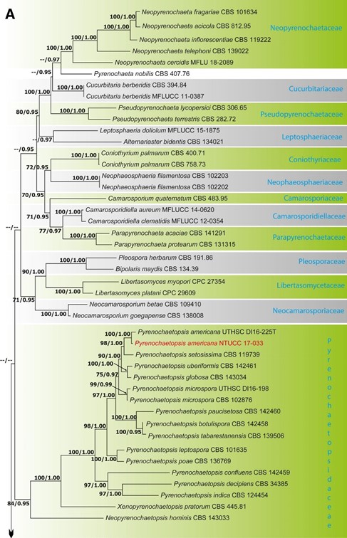
Figure 1. (A) Part one of the phylogenetic tree based on the concatenated alignment of six molecular markers (ITS, LSU, SSU, rpb2, tef1 and tub2) evaluated using RAxML. The new isolates are shown in red. ML bootstrap values (MLBS) ≥ 70% and Bayesian posterior probabilities (PP) ≥ 0.95 are presented at the nodes. The scale bar indicates the number of estimated substitutions per site. Hysterium rhizophorae (PUFD43) was used as an outgroup for rooting the tree. (B) Part two, (C) Part three, (D) Part four, (E) Part five of the phylogenetic tree based on the concatenated alignment of six molecular markers (ITS, LSU, SSU, rpb2, tef1 and tub2) evaluated using RAxML.
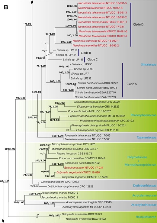
Figure 1. (continued)
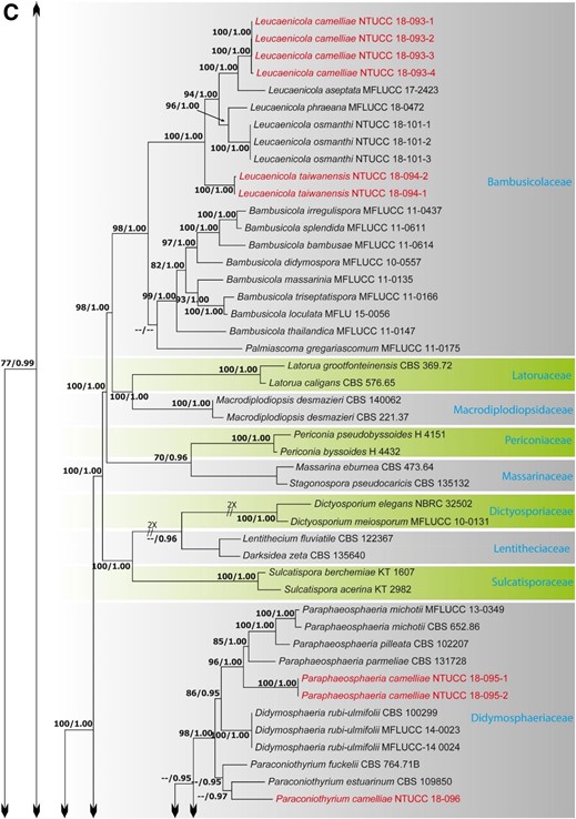
Figure 1. (continued)
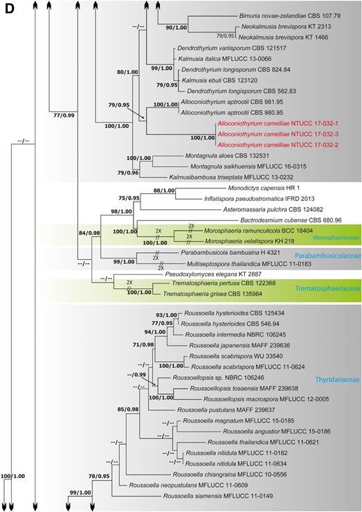
Figure 1. (continued)
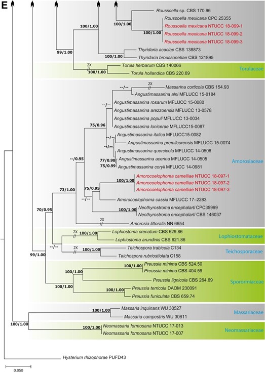
Figure 1. (continued)
Species
