Neoshiraia camelliae Ariyawansa, I. Tsai & Thambugala sp. nov.
MycoBank number: MB 834501; Index Fungorum number: IF 834501; Facesoffungi number: FoF 11720; Fig. 3.
Etymology Named after the host genus, Camellia, from which it was isolated.
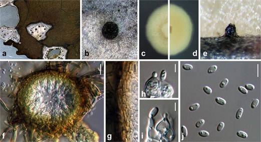
Associated with leaf lesions of C. sinensis. Leaf lesions expanded, coalesced, and irregular. Sexual morph: undetermined. Asexual morph: Conidiomata pycnidial, scattered over the surface of the leaf lesions, semi-immersed to erumpent, black, globose to subglobose, uni-loculate. Conidiomatal wall consisting of dark brown to lightly- pigmented thick-walled cells of textura angularis, 4–7 × 1–3 μm (x̄ = 5.4 ± 1.0 × 2.1 ± 0.5 μm, n = 29). Conidi- ophores reduced to conidiogenous cells. Conidiogenous cells 5–7 × 3–4 μm (x̄ = 5.1 ± 0.7 × 4.0 ± 0.4 μm, n = 28), enteroblastic, phialidic, hyaline, ampulliform to doliiform, discrete or integrated, smooth. Conidia 4–5 × 2–3 μm (x̄ = 4.3 ± 0.2 × 2.5 ± 0.1 μm, n = 30), hyaline, aseptate, obovoid to ellipsoidal, guttulate, smooth-walled.
Material examined: Taiwan, Lugu Township, Nantou County, Feng-Huang Tea Fields, on leaves of C. sinensis (Theaceae), 15 April 2018, Tsai Ichen, ST06-1 (NTUH 18-092-1, holotype), ex-type culture ST06-1 (NTUCC 18-092-1); ibid., ST06-2 (NTUH 18-092-2), living culture ST06-2 (NTUCC 18-092-2).

Figure 3. Neoshiraia camelliae (NTUH 18-092-1, holotype). (a) Leaf lesions. (b) Conidioma on host surface. (c, d) Cultures on PDA, from above and below. (e) Conidiomata on pine needle. (f) Vertical section through conidioma. (g) Conidiomatal wall. (h, i) Conidiogenous cells and developing conidia. (j) Conidia. Scale bars: (f) = 10 μm, (g, j) = 5 μm, (h, i) = 2.5 μm.
