Neopestalotiopsis mianyangensis W.L. Li & Jian K. Liu, in Li, Dissanayake, Zhang, Maharachchikumbura & Liu, Journal of Fungi 8(11, no. 1175): 10 (2022)
Index Fungorum number: IF 845406; MycoBank number: MB 845406; Facesoffungi number: FoF 12746;
Etymology – Name reflects the location Mianyang city where the holotype was collected.
Holotype – HKAS 123211
Pathogenic on diseased branches of Paeonia suffruticosa Andr. Sexual morph: Not observed. Asexual morph: Conidiomata 139–143 μm high, 245–250 μm diam. (x̄ = 141 × 248 μm, n = 15), globose, solitary, semi-immersed, black, excluding dark brown to black masses of conidia. Conidiomata wall 24–30 μm thick at sides, not well defined, comprising brown, thin-walled cells of textura angularis, with lighter cells at the base fusing into the host tissue. Conidiophores indistinct, often reduced to conidiogenous cells. Conidiogenous cells 3–5 × 2.1–2.5 μm (x̄ = 4 × 2.5 μm, n = 30), mostly integrated, ampulliform to lageniform, hyaline to light brown, smooth-walled, single, with truncated apex. Conidia 19–23 × 5.5–7 μm (x̄ = 21 × 6.5 μm, n = 30), fusoid, ellipsoid, straight to slightly curved, 4 septa; conical to obconical basal cell with a truncated base, hyaline, rough and thin-walled, 3–4 μm long, often with a small basal appendix; three medial cells with light brown to dark pigmentation with a rough wall, and one septum darker than the rest of the cell (second cell from the base pale brown, 4–5 μm; third cell golden brown 4–5.5 μm long; fourth cell brown, 4–5.5 μm long); apical cell of 3–4 μm long, hyaline, cylindrical, thin and smooth-walled with 3 apical tubular appendages, not arising from the apical crest, but each inserted at a different locus in the upper half of the apical cell, unbranched, filiform, 5.5–11 μm; single basal appendix, tubular, unbranched, centric, 3–4 μm, mean conidium length/width ratio = 3.3:1.
Culture characteristics – Colonies on PDA reaching 70 mm diam. after 7 d at 25 ℃. Colonies filamentous to circular, medium dense, with white sparse mycelium.
Material examined – China, Sichuan Province, Mianyang city, on a diseased branch of Paeonia suffruticosa, 10 September 2021, W.L. Li, YMD 451 (HKAS123211, holotype), ex-type living culture, CGMCC3.23554; ibid., HUEST 22.0066, isotype, ex-isotype living culture, UESTCC 22.0066.
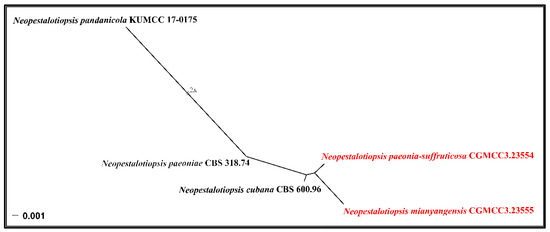
Notes – Neopestalotiopsis mianyangensis (UESTCC 22.0006) forms a sister clade to N. cubana (CBS 600.96), N. pandanicola (KUMCC 17–0175), and N. paeoniae (CBS 318.74) in a poorly-supported clade (Figure 2). Neopestalotiopsis mianyangensis differs from N. cubana by its shorter conidiogenous cells (3–5 μm vs. 5–12 μm) and shorter apical appendages (5.5–11 μm vs. 21–27 μm), as well as narrower conidia (5.5–7 μm vs. 8–9.5 μm). Neopestalotiopsis pandanicola can be morphologically distinguished from N. mianyangensis by larger conidia (27–35 μm vs. 19–23 μm) and longer appendages (9.5–26 μm vs. 5.5–11 μm). Neopestalotiopsis paeoniae was originally described from Anacardium occidentale [19]. Unfortunately, no living culture was obtained for this species, and only ITS and tef1-α sequences are available. Neopestalotiopsis mianyangensis and N. paeoniae had seven base pairs differences in tef1-α loci without gaps. The PHI analysis further confirms that N. mianyangensis has no significant genetic recombination with closely related species (Fw > 0.05, Figure 6). We thus introduced N. mianyangensis as a new species.

Figure 1 – Neopestalotiopsis mianyangensis (HKAS 123211, holotype). a, b Appearance of conidiomata on the host. c Transverse section of conidiomata. d Conidiomatal wall. e Sporodochia. f, g Conidiogenous cells and conidia. h–n conidia. o Germinating conidium. Scale bars: c = 50 μm, d–o = 10 μm.