Neomassaria alstoniae N.I. de Silva, Lumyong & K.D. Hyde, sp. nov.
MycoBank number: MB 559519; Index Fungorum number: IF 559519; Facesoffungi number: FoF 10717; Fig. 6.22
Etymology: Name reflects the host genus Alstonia, from which the new species was isolated.
Holotype: MFLU 21-0238.
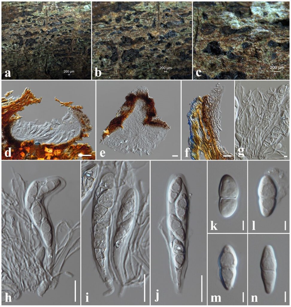
Saprobic on dead twigs attached to Alstonia scholaris. Sexual morph: Ascomata 170–300 μm high × 300–350 μm diam. ( = 250 × 320 µm, n = 10), solitary or scattered, coriaceous, immersed to slightly erumpent, visible as black dots on the host surface, unilocular, globose to subglobose, brown to dark brown. Ostiole central. Peridium 20–36 μm wide, comprising light brown cells of textura angularis, cells towards the inside hyaline to lightly pigmented, fusing at the outside indistinguishable from the host tissues. Hamathecium comprising 1–2 μm wide, cylindrical to filiform, septate, branched, pseudoparaphyses. Asci 80–100 × 12–18 μm (= 92 × 16 μm, n = 20), 8-spored, bitunicate, oblong to cylindrical, short pedicellate, with ocular chamber. Ascospores 20–24 × 7–10 μm (= 22 × 8.5 μm, n = 30), overlapping 1–2-seriate, hyaline, ellipsoid to fusiform, 1-septate, constricted at the septum, without a mucilaginous sheath. Asexual morph: Undetermined.
Culture characteristics – Colonies on PDA reaching 25 mm diameter after 2 weeks at 25°C, colonies from above: white, irregular, undulate margin, flat, slightly raised, fluffy appearance, cream at the margin; reverse: brown from the centre of the colony, cream at margin.
Material examined – THAILAND, Chiang Rai Province, dead twigs attached to Alstonia scholaris (Apocynaceae), 25 April 2019, N. I. de Silva, AS14 (MFLU 21-0238, holotype), ex-type living culture, MFLUCC 21-0213.
Notes – Neomassaria alstoniae was collected from dead twigs of Alstonia scholaris in Thailand. According to the multi-gene phylogeny, Neomassaria alstoniae forms a sister lineage to N. formosana with 94% ML and 1.00 BYPP support (Fig. **). Neomassaria formosana can be distinguished from N. alstoniae in having distinct neck their ascomata and periphyses (Ariyawansa et al. 2018). Neomassaria formosana was introduced by Ariyawansa et al. (2018) from a dead stem of Rhododendron species in Taiwan. Additional morphological differences are mentioned in Table 3.

Figure 6.22 Neomassaria alstoniae (MFLU 21-0238, holotype). a–c Appearance of ascomata on substrate. d Vertical sections through ascoma. e Vertical sections through neck region. f Peridium. g Pseudoparaphyses. h–j Asci. k–n Ascospores. Scale bars: a–c = 200 μm, d = 50 μm, e, f = 20 μm, g = 5 μm, h–j = 20 μm, k–n = 5 μm.
