Pseudochaetosphaeronema magnoliae N.I. de Silva, Lumyong & K.D. Hyde, sp. nov.
MycoBank number: MB 559518; Index Fungorum number: IF 559518; Facesoffungi number: FoF 10716; Fig. 6.18
Etymology: Name reflects the host genus Magnolia, from which the new species was isolated.
Holotype: MFLU 18-1296
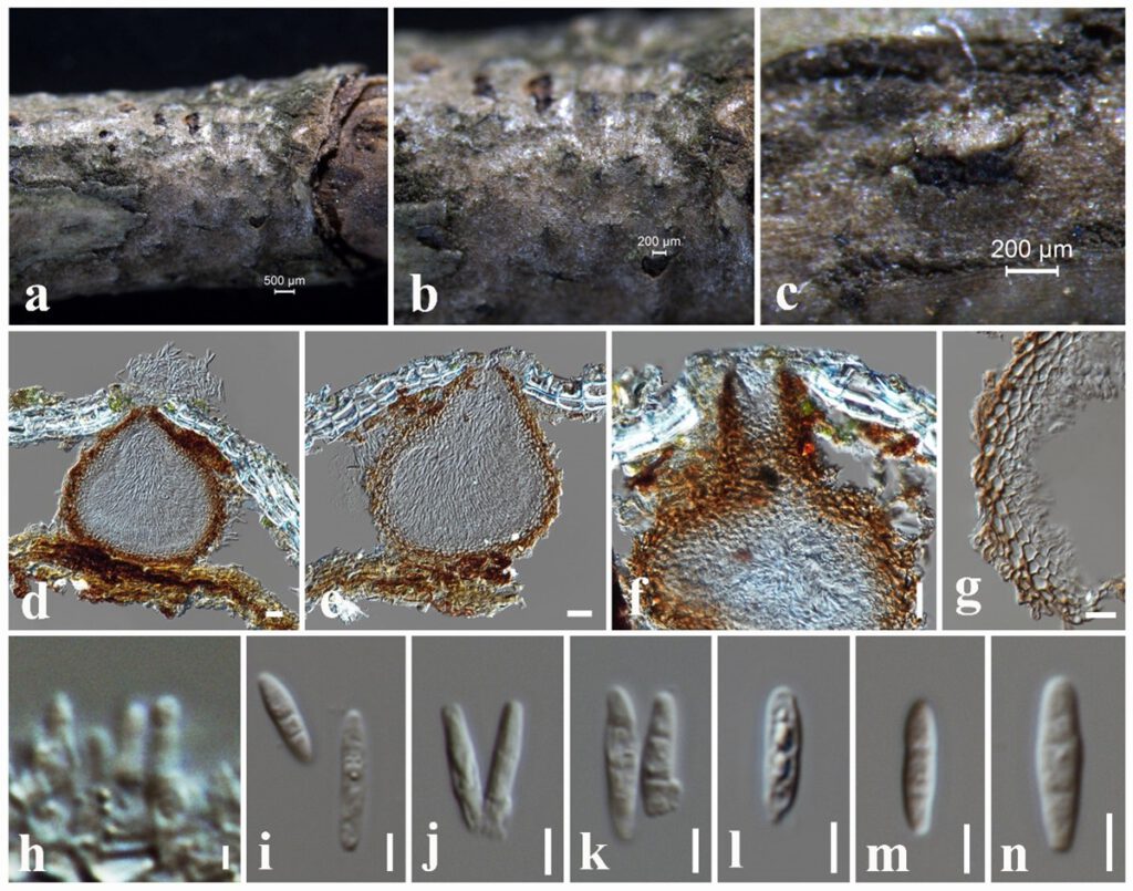
Saprobic on dead twigs attached to Magnolia garrettii. Sexual morph: Undetermined. Asexual morph: Conidiomata 160–190 × 150–180 µm ( = 170 × 165 μm, n = 10), solitary, globose to subglobose, dark brown to black, immersed to erumpent, solitary, unilocular, ostiolate. Conidiomatal wall 15–20 μm wide, composed of several layers of small, flattened, brown to dark brown pseudoparenchymatous cells, cells towards the inside lightly pigmented, arranged in a textura angularis, at the outside, darker, fusing and indistinguishable from the host tissues. Conidiophores reduced to conidiogenous cells. Conidiogenous cells 4–5 × 2.5–3.5 µm ( = 4.6 × 3.2 μm, n = 20), produced from inner stromatic tissue, monophialidic, cylindrical or ampulliform, integrated, hyaline, smooth-walled. Conidia 12–18 × 2–3.5 μm ( = 14 × 3 μm, n = 40), solitary, hyaline, smooth, thin-walled, straight, cylindrical to fusoid, apex obtuse, unicellular, without mucilaginous sheath.
Culture characteristics – Colonies on PDA reaching 30 mm diameter after 1 weeks at 25°C, colonies from above: circular, margin entire, dense, slightly raised, white at the margin, light brown in the centre; reverse: cream at the margin, dark brown in the centre.
Material examined – THAILAND, Chiang Mai Province, dead twigs attached to host plant of Magnolia garrettii (Magnoliaceae), 13 September 2017, N. I. de Silva, NI197 (MFLU 18-1296, holotype), ex-type living culture, MFLUCC 18-0707, CHINA, Yunnan Province, Xishuangbanna, dead twigs attached to the host plant of Magnolia liliifera (Magnoliaceae), 27 March 2017, N. I. de Silva, NI167 (MFLU 18-1028), living culture, KUMCC 17-0196.
Notes – According to the multi-gene phylogeny herein, Pseudochaetosphaeronema magnoliae clustered with P. kunmingense and P. siamense with 100% ML and 1.00 BYPP support. Pseudochaetosphaeronema magnoliae can be distinguished from P. kunmingense in having smaller conidiomata (160–190 × 150–180 µm vs 180–250 μm diam.) and hyaline, aseptate conidia (12–18 × 2–3.5 μm), whereas P. kunmingense has light brown, 3-septate conidia (10–15 × 4–6 μm) (Hyde et al. 2020). In addition, Pseudochaetosphaeronema magnoliae differs from P. siamense from distinct size differences of conidiomata (160–190 × 150–180 µm vs 85–100 × 80–90 µm), conidiogenous cells (4–5 × 2.5–3.5 µm vs 8–17 × 1–2.5 µm) and conidia (12–18 × 2–3.5 μm vs 3–5 × 2.5–3 µm) (Jayasiri et al. 2019). It is interesting to note that Pseudochaetosphaeronema magnoliae recorded from both China and Thailand.

Figure 6.18 Pseudochaetosphaeronema magnoliae (MFLU 18-1296, holotype) a The specimen. b, c Appearance of immersed conidiomata on substrate. d, e Vertical sections through conidiomata. f Vertical sections through conidiomata and neck region. g Conidiomatal wall. h Conidiogenous cells. i–n Conidia. Scale bars: a = 500 μm, b, c = 200 μm, d–f = 20 μm, g = 10 μm, h = 2 μm, i–n = 5 μm.
