Neogloeosporidina pruni W.J. Li, Camporesi & K.D. Hyde, sp. nov.
MycoBank number: MB 557153; Index Fungorum number: IF 557153; Facesoffungi number: FoF 07476; Fig. 251
Etymology: Named after the host from which it was collected, Prunus.
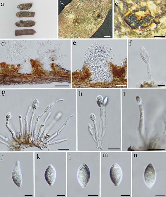
Saprobic on dead stems of Prunus avium (Rosaceae). Sexual morph: undetermined. Asexual morph: Conidiomata 100–400 µm diam., 90–230 µm high, brown, eustromatic, pycnidial, scattered, rarely gregarious, subepidermal or subperidermal in origin, immersed to partly erumpent, globose to subglobose, unilocular, thick-walled, smooth, glabrous. Ostiole absent, dehiscing by an irregular rupture in the apical wall, then becoming wide open. Conidiomata wall 20–30 µm wide, composed of brown, thick-walled cells of compressed textura angularis, Conidiophores mostly reduced to conidiogenous cells, occasionally present, short, cylindrical, septate and branched at base, formed from inner layers of conidiomata. Conidiogenous cells 10–35 × 2.1–3.9 µm, hyaline, enteroblastic, cylindrical, discrete or integrated, indeterminate, smooth-walled. Conidia 11–18 × 4.5–9 µm ( x̄ = 14 × 6.9 µm; n = 30), hyaline, ellipsoid to ventricose, often with a blunt apiculus, unicellular, thick-walled, smooth.
Material examined: Italy, Province of Forlì-Cesena, Santa Sofia, Campigna, on dead land branches of Prunus avium (Rosaceae), 26 July 2014, Erio Camporesi, IT2016 (MFLU 16-2153, holotype), (KUN, HKAS 101641, isotype).

Fig. 251 Neogloeosporidina pruni (MFLU 16-2153, holotype) a Herbarium specimen. b, c Appearance of black conidiomata on the host. d, e Vertical sections of conidiomata. f–i Conidiophores, conidiogenous cells and developing conidia. j–n Conidia. Scale bars b, d = 100 µm, c = 200 µm, e = 50 µm, f, j–n = 5 µm, g–i = 10 µm
