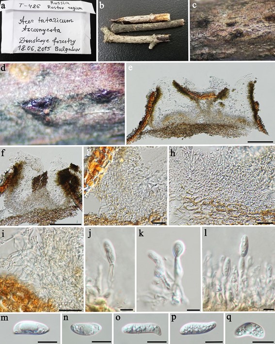
Neodermea rossica W.J. Li, D.J. Bhat & K.D. Hyde, sp. nov.
MycoBank number: MB 557152;Index Fungorum number: IF 557152; Facesoffungi number: FoF 07473; Fig. 243
Etymology: Named after country Russia from which it was collected.
Saprophytic on a dead stem of Acer tataricum (Sapindaceae). Sexual morph: undetermined. Asexual morph: Conidiomata 370–450 µm diam., 200–350 µm high, dark brown to black, stomatic, pycnidial, solitary or gregarious, deeply immersed in origin, becoming partly erumpent at maturity, globose to subglobose, unilocular or multi-locular, thick-walled, ostiolate. Ostiole cylindrical, single, with a wide channel, centrally located. Conidiomata wall 30–90 µm wide, composed of hyaline to brown, rather thick-walled cells of textura angularis to textura prismatica at basal region, and brown to dark brown cells of textura angularis at upper and lateral region. Conidiophores hyaline, cylindrical, septate, arising from the inner layers of conidiomata. Conidiogenous cells 3–15 × 2–4 µm, enteroblastic, phialidic, cylindrical, discrete or integrated, indeterminate, smooth. Conidia 14–25 × 4–8 µm (x̄ = 17 × 6.5 µm; n = 30), hyaline, oblong-ellipsoid, rounded at both ends, or narrowed and truncate at base, straight or slightly curved, unicellular, guttulate, smooth. Microconidia absent.
Material examined: Russia, Rostov region, Donskoye forestry, on dead stems of Acer tataricum (Sapindaceae), 18 June 2015, T.S. Bulgakov, T486B (MFLU 15-2190, holo-type), ex-type living culture MFLUCC 17-2506 = KUMCC 15-0635, T486 (MFLU 19-2890).
Notes: Neodermea rossica is similar to N. acerina in its oblong-ellipsoid, straight or slightly curved, unicelluar conidia. However, those two taxa are separated by conidiomatal structure and microconidia. Neodermea acerina has slender, beaked conidiomata up to 1.5 mm in length and 300–650 µm wide, which is longer and wider than the globose to subglobose conidiomata with cylindrical ostiole in N. rossica (Grove 1946). Neodermea acerina also has micro-conidia, which are absent in N. rossica.

Fig. 243 Neodermea rossica (MFLU 15-2190, holotype). a, b Her- barium specimen. c, d Appearance of black conidiomata on the host. e, f Vertical sections of conidiomata. g–i Section of peridium. j–l Conidiophores, conidiogenous cells and developing conidia. m–q Conidia. Scale bars e–f = 200 µm, g–i = 20 µm, j–l = 5 µm, m– q = 10 µm
