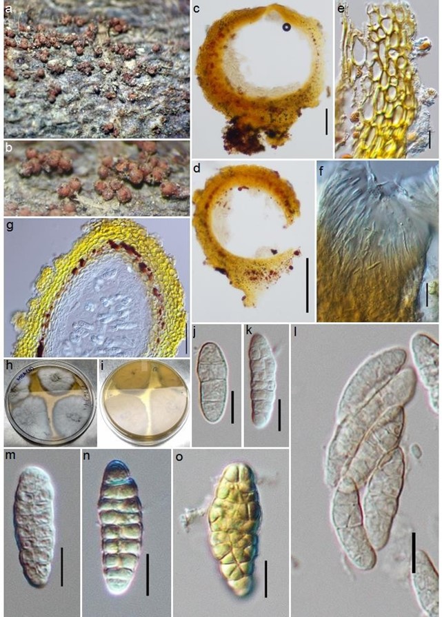
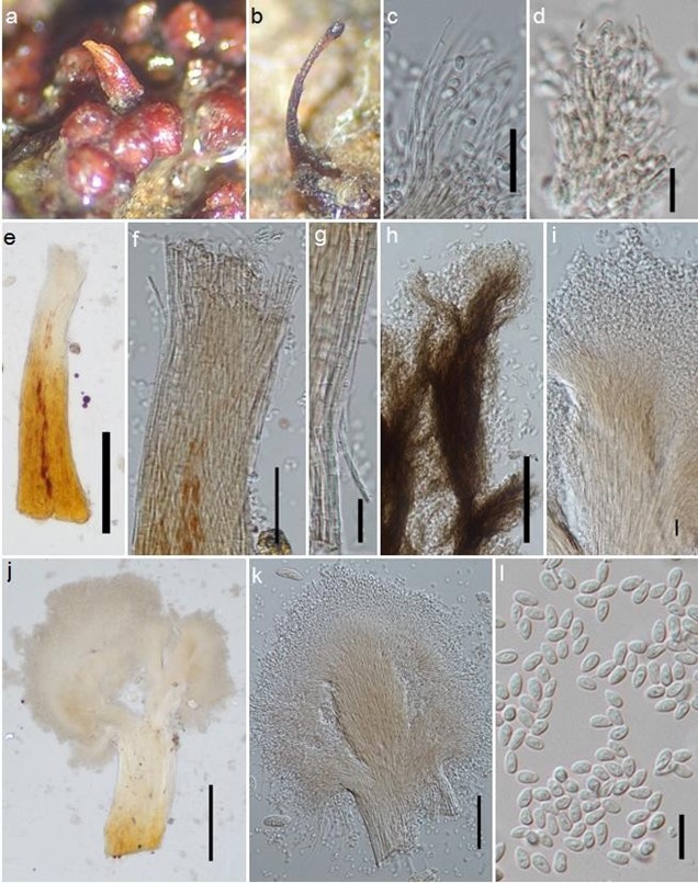
Murinectria murispora M. Niranjan & V.V. Sarma, sp. nov. Figs 175, 176
MycoBank number: MB 556613; Index Fungorum number: IF 556613; Facesoffungi number: FoF 06267;
Etymology – The specific epithet “murispora” refers to the fungus having muriform ascospores.
Holotype – AMH-10077.
Saprobic on decaying climber. Sexual morph: Stromata up to 1 mm high and 3.2 mm diam., erumpent through epidermis, subiculate, pseudoparenchymatous cells forming textura prismatica cell layers, intergrading with peridium. Ascomata 550–580 × 415–500 μm, perithecial, globose, superficial, gregarious, KOH+ dark red, LA+ yellow, often associated with synnemata of the asexual state, depressed apical region, periphysate, apical region without stroma. Peridium up to 43 μm thick, wall consists of textura globulosa and textura angularis cells, walls pigmented. Asci 64– 92 × 14.5–18 μm, 8-spored, unitunicate, cylindric-clavate, with an inconspicuous ring at apex, early deliquescent. Ascospores (25–) 26–36.6 (–38) × 10 – 16.5 (–18.5) μm ( x = 32.8 × 14.9, n = 28), overlapping biseriate, hyaline, yellow in Lugol’s solution, ellipsoidal to fusiform, muriform, with 5–9 transverse septa and 1–5 longitudinal septa, often constricted at each septum, straight, sometimes slightly curved, smooth-walled. Asexual morph: Synnematous on natural substrata, 892 μm length × 144–164 μm diameter, laterally oriented near perithecial ascomata, usually erumpent through epidermis, solitary to gregarious around ascomata, erect or nodding, unbranched, narrowing towards apex, red-brown at base, becoming dark brown to black with age, ovoid heads consisting of pools of conidia, individual conidiophores septate. Conidiogenous cells phialidic. Conidia 4.7–6.3 × 2.6–3.4 μm ( x = 5.5 × 3, n = 28), hyaline, obovoid, slightly flat to rounded apex, narrow towards base, acute ends, smooth-walled.
Culture characteristics – White cottony colonies on malt extract agar, becoming gray-brown at maturity, filamentous, radial, background pale yellow colour, 42 mm diameter in one-week old culture grown at 28 °C.
Material examined – INDIA, Andaman and Nicobar Islands, South Andaman, Pongibalu, Manjery (11˚52’25.7”N 92˚64’89.9”E), on decaying twig, 10 December, 2017. M. Niranjan PUFNI 17634 (AMH-10077, holotype), extype-living culture, NFCCI-4515.
GenBank numbers – ITS: MK860769, LSU: MK860767.
Notes – Murinectria murispora has similar characters to three species of Nectria in having muriform ascospores (Table 1) (Hirooka et al. 2012). In comparison to Murinectria murispora, Nectria polythalama stromata are shorter and narrower and have smaller ascomata and ascospores. The synnemata (asexual state) on natural substrata are scattered or grow around the stromata of Murinectria murispora, while in Nectria polythalama, the synnemata are frequently found in the middle of the stromata. Nectria pseudotrichia also resembles Murinectria murispora in having similar ascomata and muriform ascospores, but is distinct in having smaller ascomata and ascospores. Nectria antarctica also has muriform ascospores. The ascostromata and ascospores of N. antarctica, however, are smaller and have longer asci when compared to Murinectria murispora. Murinectria murispora is quite similar to Nectria polythalama and N. pseudotrichia in terms of septal constrictions, unlike N. antarctica. Murinectria murispora is distinct from all three species in often having 5 longitudinal septa. Hence, based on the above mentioned differences and DNA sequence analysis, a new species, M. murispora is introduced.

Figure 175 – Murinectria murispora (AMH-10077, holotype). a, b Ascomata on host. c, d, g Vertical section of ascomata. e Peridium. f Neck. h, i Culture on MEA plate. l Asci. j, k, m-o Ascospores. Scale bars: d = 100 μm, d, f, g = 50 μm, j-o = 10 μm.

Figure 176 – Murinectria murispora (AMH 10077). a, b Synnemata on natural substrate. c-e Synnemata. f, g Synnemata head. h, i Conidiophores and conidia. j, k Conidiogenous cells. l Conidia. Scale bars: f = 200 μm, c, d, g, h = 50 μm, e, j, k 20 μm, i, l = 10 μm.
