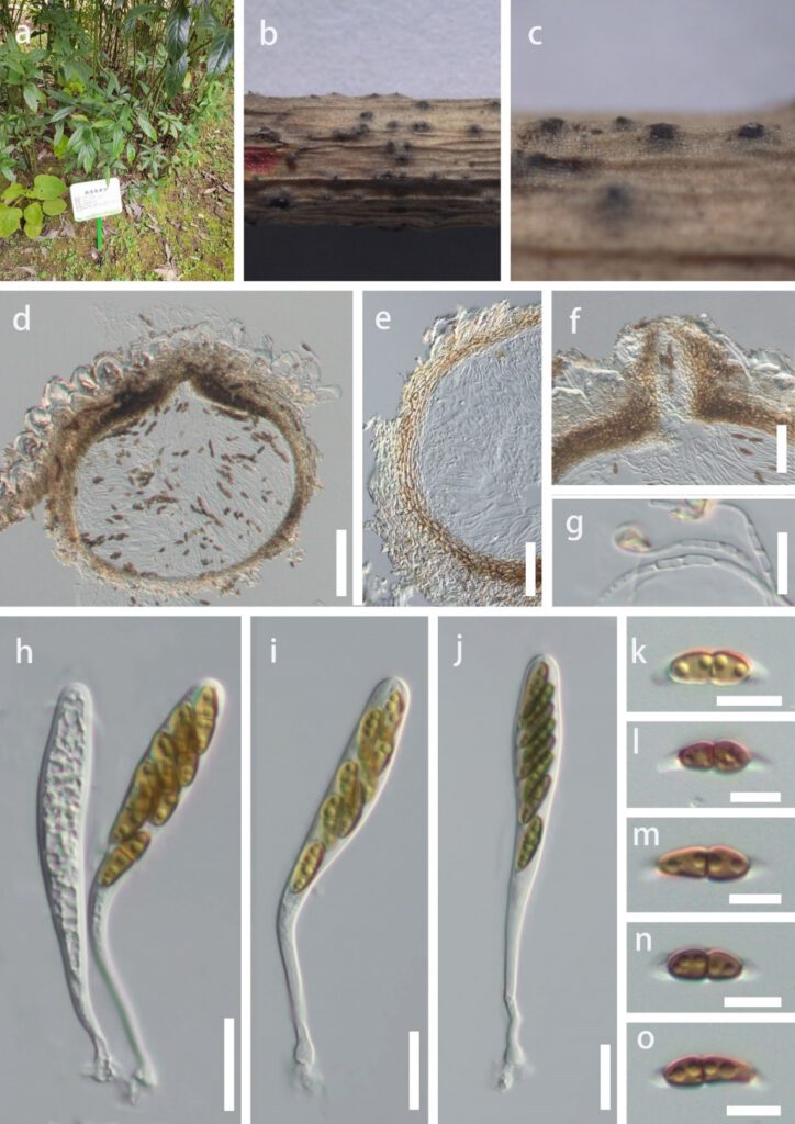
Montagnula guiyangensisY.R. Sun, Yong Wang bis & K.D. Hyde, sp. nov. (Fig.3)
MycoBank number: MB; Index Fungorum number: IF; Facesoffungi number: FoF 12923;
Holotype: HKAS 124556
Etymology: Referring to the location in which the fungus was collected.
Saprobic on twigs of Helwingia himalaica in terrestrial habitat. Sexual morph: Ascomata 300–400 × 350–400 μm, semi-immersed, solitary or scattered, globose, uniloculate, black, with a central ostiole. Ostiole papillate, central. Peridium 20–40 μm wide, fused with host tissues, comprising of 2 layers of pale brown to brown cells of textura angularis. Hamathecium comprises 1.5–3.0 μm wide, branched, hyaline, septate, pseudoparaphyses. Asci 84–135 × 10–15 μm ( = 104 × 12 μm, n = 15), bitunicate, 8-spored, clavate, with a short, bulbous long pedicel, slightly curved. Ascospores 10–20 × 3.5–6 μm ( = 15.5 ×5 μm, n = 35), hyaline to olivaceous when immature, brown when mature, overlapping uniseriate or 2-seriate, fusiform, 1-septate, constricted at the septum, slightly widest at the upper cell and tapering towards ends, guttulate, sheath drawn out to form polar appendages, from both ends of the ascospores, straight or slightly curved. Asexual morph: Not observed.
Culture characteristics: Ascospores germinating on PDA within 12 h at 25 ˚C, germ tubes produced from both ends. Colonies on PDA reaching 7 cm diam after 4 weeks at 25 ˚C, mycelium white to gray, flossy, circular, slightly flattened, undulate, yellow in reverse.
Material examined: China, Guizhou Province, Guiyang City, Nanming District, Guiyang Medicinal Botanical Garden, on twigs of Helwingia himalaica, 22 December 2021, Y.R. Sun, 22-41 (HKAS 124556, holotype; ex-type living culture GUCC 816); ibid, on twigs of Helwingia himalaica, 22 December 2021, Y.R. Sun, 41-2 (HGUP 800, living culture GUCC 817).
Notes: Montagnula guiyangensis was isolated from Helwingia himalaica, an important medicinal plant. Multi-gene analyses showed that M. guiyangensis is a phylogenetically distinct species in Clade 2 (Fig. 1). Morphologically, M. guiyangensis resembles M. appendiculata, M. chiangraiensis and M. chromolaenae in having fusiform, 1-septate ascospores with appendages. But M. guiyangensis differs by larger ascomata from M. chromolaenae and M. appendiculata (300–400 × 350–400 μm in M. guiyangensis vs. 170–190 × 170–190 μm in M. chromolaenae vs. 100–200 μm in M. appendiculata) (Mapook et al. 2020). And M. guiyangensis has larger asci than M. chiangraiensis (84–135 × 10–15 μm vs. 60– 75 × 8–11). Montagnula guiyangensis can be distinguished from M. aloes by 1-septate, fusiform ascospores with appendages, while the latter has 3-euseptate, ovoid to ellipsoid ascospores(Crous et al. 2012). In addition, comparisons of ITS, LSU and SSU sequences between M. guiyangensis and phylogenetically related species are provided (Table 4).
Table 4. Number of polymorphic nucleotide differences between M. guiyangensis HKAS 124556 with M. aloes, M. appendiculata, M. chiangraiensis and M. chromolaena (without gap).
| Species | Strain | ITS | LSU | SSU |
| M. aloes | CBS 132531 | 20(497bp) | 10(815bp) | not available |
| M. appendiculata | CBS 10927 | 18(497bp) | 16(813bp) | not available |
| M. chiangraiensis | MFLUCC 17-1420 | 13(497bp) | 18(813bp) | 27(987bp) |
| M. chromolaena | MFLUCC 17-1435 | 14(497bp) | 18(813bp) | 32(987bp) |

Figure 3. Montagnula guiyangensis (HKAS 124556, holotype). a. Host. b, c. Appearance of ascomata on the substrate. d. Section through ascomata. e. Peridium. g. Trabeculate pseudoparaphyses. h–j. Asci. k–o. Ascospores. Scale bars: d = 100 μm, e, f = 50 μm, g–j = 20 μm, k–o = 10 μm.
