Micromelanconis kaihuiae C.M. Tian & N. Jiang, sp. nov.
MycoBank number: MB 838928; Index Fungorum number: IF 838928; Facesoffungi number: FoF; Figures 2, 3
Etymology. Named after Kaihui Yang, a Chinese heroine; Kaihui is also the name of the town where holotype was collected.
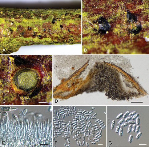
Description. Sexual morph: not observed. Asexual morph: Conidiomata acervular, 350–800 μm diam., conspicuous, immersed in host bark to erumpent, covered by brown to blackish exuding conidial masses at maturity. Central column beneath the disc more or less conical. Conidiophores unbranched, aseptate, cylindrical, pale brown, smooth-walled. Conidiogenous cells annellidic, occasionally with distinct annellations and collarettes, 12.4–47.1 × 1.2–3.8 μm. Conidia hyaline when immature, becoming pale brown, ellipsoid, multiguttulate, aseptate, 7.6–10.3 × 3.1–4.1 μm, L/W = 2–3.2, with hyaline sheath, 1 μm.
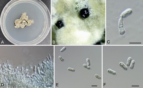
Culture characters. Colony on PDA at 25 °C irregular, grey olivaceous, margin becoming diffuse, aerial hyphae short, dense, surface becoming imbricate, growth lim- ited and ceasing after two weeks. Conidiomata formed after three weeks, randomly distributed, black. Conidiophores unbranched, septate, cylindrical, pale brown, smooth- walled. Conidiogenous cells annellidic, 9.1–18.5 × 2.5–5.3 μm. Conidia pale brown, long dumbbell-shaped, narrow at the middle and wide at both ends, multiguttulate, aseptate, 10.4–13.5 × 4–5 μm, L/W = 2.3–3.3, with hyaline sheath, 1.5 μm.
Specimens examined. China, Hunan Province, Changsha City, Changsha Coun- ty, Kaihui Town, chestnut plantation, 40°24’32.16″N, 117°28’56.24″E, 262 m asl, on stems and branches of Castanea mollissima, Tian Chengming and Ning Jiang, 10 November 2020 (BJFC-S1831, holotype; ex-type culture, CFCC 54572 = KH5-3). Ibid. (BJFC-S1832, KH5-4).
Notes. Micromelanconis kaihuiae on Castanea mollissima (Fagaceae, Fagales) is phylogenetically close to Neopseudomelanconis castaneae on Castanea mollissima and Pseudomelanconis caryae on Carya cathayensis (Juglandaceae, Juglandales) (Fig. 1). All these three species are discovered on tree branches in China, and share similar morphological characters in having pale brown conidia with conspicuous hyaline sheath. Micromelanconis kaihuiae and Neopseudomelanconis castaneae even share the same host. However, they can be easily distinguished based on conidia shape, color and overall size of conidia (M. kaihuiae, pale brown, ellipsoid and aseptate conidia, 7.6–10.3 × 3.1–4.1 μm; pale brown, long dumbbell-shaped and aseptate conidia, 10.4–13.5 × 4–5 μm vs. N. castaneae, brown, ellipsoid to oblong and septate conidia, 18–21.5 × 4.8–7 μm vs. P. caryae, pale brown, ellipsoid to oblong and aseptate conidia, 12.5–16 × 4–5 μm) (Fan et al. 2018a; Jiang et al. 2018a). Furthermore, M. kaihuiae is separated from N. castaneae by 51/490 bp (10.4%) differences in ITS and 12/563 bp (2.1%) differences in LSU, and from P. caryae by 56/490 bp (11.4%) differences in ITS and 6/563 bp (1.1%) differences in LSU.

Figure 2. Morphology of Micromelanconis kaihuiae on branches of Castanea mollissima (BJFC-S1831) A, B habit of conidiomata on a branch C transverse section of conidiomata D longitudinal section through co- nidiomata E conidiogenous cells attached with conidia F, G conidia. Scale bars: 100 μm (C, D); 10 μm (E–G).

Figure 3. Morphology of Micromelanconis kaihuiae on the PDA plate (CFCC 54572) A colony on PDA B habit of conidiomata formed on PDA C, D conidiogenous cells attached with conidia E, F conidia. Scale bars: 10 μm (C–F).
