Meliola tamarindi Syd. & P. Syd., Annls mycol. 10(1): 79 (1912)
Index Fungorum number: IF247233, Facesoffungi number: FoF00489, Fig. 2
Epifoliar pathogen on living leaves. Sexual morph Colonies epiphyllous, scattered, 1-2 mm diam. Hyphae superficial, substraight to crooked, branching opposite at acute to wide angles, closely reticulate, with dark brown mycelial setae. Appressoria 20–25×9–12μm (x=22×10μm, n=10), opposite to alternate, straight to curved, two-celled, clavate to spathulate. Phialides 20–22(-25)×7–9μm (x=22×8μm, n= 5), few mixed with appressoria, alternate to opposite, ampulliform. Ascomata 210–240μm wide (x=222μm, n= 10) and 170–220μm high (x=195μm, n=10), subdense, superficial on the mycelium, composed of hyaline inner cell and dark brown outer wall with textura angularis, globose to subglobose, dark brown, with ostiolate and dark brown setae. Asci 65–85×28–40μm (x=73×34μm, n=5), 2-spored, unitunicate, ellipsoid to ovoid, evanescent. Ascospores 43–48× 19–22μm (x=45×20μm, n=20), ellipsoid to fusiform, hyaline with 2 septa when young state, becoming dark brown with 4 septa when mature, constricted at septa, middle cell slightly longer than others. Asexual morph Undetermined.
Material examined: THAILAND, Chiang Rai province, Bandu; on the living leaves of Tamarindus indica L.=(Fabaceae), 10 January 2014, Xiang-Yu Zeng (MFLU 14– 0282, reference specimen designated here). GenBank LSU: KP744489.
Notes: In Fig. 1, Meliola tamarindi clusters with a putatively named strain of Asteridiella obesa and four other Meliola species. Asteridiella sp., Endomeliola dingleyae and Appendiculella sp. also cluster in Meliolaceae (Fig. 1). Our collection of Meliola tamarindi has larger spores than in the protologue (43–48×19–22μm, versus 36–44×13–17μm (Sydow and Sydow 1912). The addition of reference specimen sequence data for M. tamarindi is important for this phylogenetically poorly understood group.
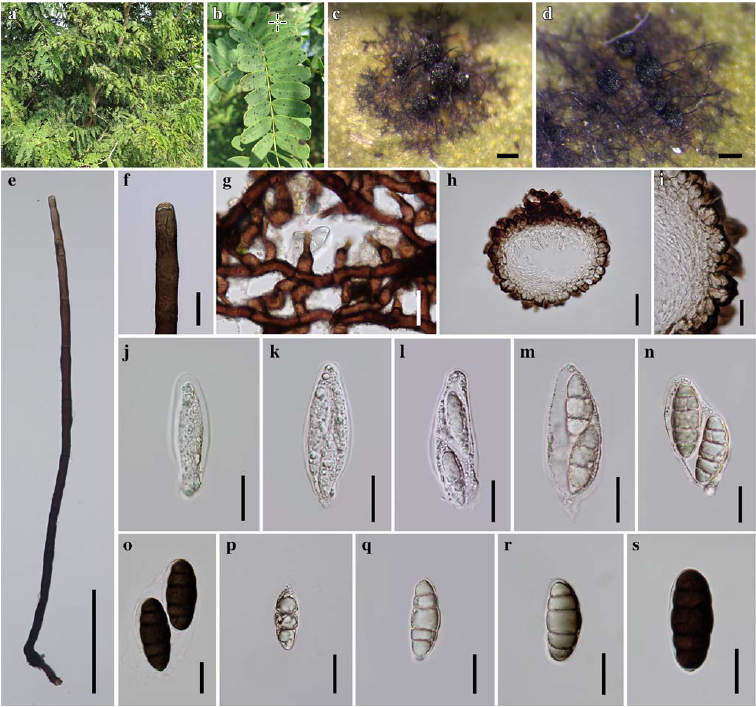
Fig. 1 Meliola tamarindi (MFLU 14–0282). a Host b Host leaves c Colony on leaf d Ascomata e Seta f Apex of seta g Mycelium with phialides and appressoria h Cross section of ascoma I Peridium, j–o Immature and mature asci, p–s Immature and mature ascospores. Scale bars: c-d=200μm, e=100μm, h-i=50μm, f-g, j-s=20μm.

Fig. 2 Phylogram generated from Bayesian analysis based on LSU sequence data of Meliolaceae. Parsimony bootstrap support values greater than 50 % are indicated above the nodes, and branches with Bayesian posterior probabilities greater than 0.95 are given in bold. The ex-types (ex-epitypes, reference strains) are in bold, the new isolates are in blue. The tree is rooted with Chaetomium globosum CBS 147.51.