Hapalocystis berkeleyi Auersw. ex Fuckel, Fungirhenani exsic., fasc. 6: no. 585 (1863)
Index Fungorum number: IF357284, Faces of fungi number: FoF00413; Figs. 2 and 3
Reference specimen: MFLU14–0798
Saprobic on bark of Platanus hybrid. Sexual morph Ascomata 90–185μm high×206–302μm wide (x=150× 245μm, n=15), immersed, solitary or aggregated, scattered, visible through dark cracks on the host surface, subglobose to globose, ostiolate. Ostiole 130–195μm high×90–113μm wide (x=145×105μm, n=10), short papillate, black, cylindrical, periphysate. Periphyses hyaline. Peridium 4–16μm wide (x=10μm, n=15) black, thick-walled, 3–5-layered of cells of textura angularis. Paraphyses 3–7μm wide, few, septate, hyaline, attached to the base, longer than asci. Asci 112–127× 46–80μm (x=121×66μm, n=20) 4 or 8-spored, unitunicate, clavate to fusoid, with a short pedicel, with an inconspicuous flat, refractive ring at the lower end of the thickened apical wall, apex narrow and blunted. Ascospores 34–42×12–18μm (x=37×17μm) inequilaterally ellipsoidal, with broadly rounded ends, initially hyaline, dark brown at maturity, (1–)2(–3)- septate, with cells of equal length, thick and smooth-walled, with short, hyaline strap-like appendages situated at both rounded ends with same width as the ascospore. Asexual morph Conidiomata on MEA 165–195μm diam. (x= 182μm, n=15), pycnidia, superficial, aggregated, 3–5 in one group, globose, orange to brown, Conidiomatal wall 7–13μm wide (x=10μm, n=10) small, thick-walled, orange, 5–10 cells layers of textura angularis. Conidiophores 1.5–2μm high×1– 1.5μm wide (x=2×1.5μm, n=10) branched, hyaline, short, few conidiogenous cells arising from one conidiophore, attached to conidiomatal wall. Conidiogenous cells 10–14μm long, 5–7μm wide, cylindrical, hyaline, bottle-shaped, septate, ends pointed, phialidic. Conidia 0.5–1.5 diam. (x= 1μm, n=20) ellipsoid, one-celled, hyaline, smooth-walled.
Culture characters: Ascospores germinating on MEA after 2 days at 16 °C forming hyaline, branched germ tubes from end cells. Colonies on MEA reaching 2 cm after 14 days at 25 °C in light, greenishash, effuse, circular, margin entire, surface rough.
Material examined: ITALY, Province of Forlì-Cesena [FC], Modigliana, Montebello, on branch of Platanus hybrid, 14 April 2013, E. Camporesi It 1187 (MFLU 14-0798, reference specimen of Hapalocystis berkeleyi designated here); extype living cultures, MFLUCC 13-0662. GenBank LSU: KP744486.
Notes: Hapalocystis (Sydowiellaceae, Diaporthales) is typified by H. berkeleyi. LSU gene analysis of diaporthalean fungi showed Hapalocystis as a distinct genus in Sydowiellaceae (Castlebury et al. 2002). Jaklitsch and Voglmayr (2004) evaluated Hapalocystis introducing a new species. Hapalocystis comprises H. occidentalis, H. ulmi, H. bicaudata, H. corticalis and H. berkeleyi (H. berkeleyi Auersw. ex Fuckel var. berkeleyi and H. berkeleyi var. kickxii (Westend.) M.E. Barr (Jaklitsch and Voglmayr 2004). The asexual morph of Hapalocystis is given as Stilbospora, Hendersonia or Dothiorella (Wehmeyer 1941), or as stilbospora-like (Barr 1978). Glawe (1985) reported a Phoma-like asexual morph for H. berkeleyi in culture. In this study we observed a phoma-like asexual morph on MEA and we made fresh collections of Hapalocystis berkeleyi which is identical to the morphology of type specimen. The type specimen is in good condition (at herbarium K) and hence, we designate this as a reference specimen (Ariyawansa et al. 2014a) and provide molecular data; the asexual morph was also produced in culture. LSU gene analysis of taxa in family Sydowiellaceae showed our reference strain of H. berkeleyi cluster with other H. berkeleyi strains with 100 % bootstrap support (Fig. 1).
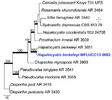
Fig. 1 Phylogram generated fromMaximum Parsimony analysis based on LSU sequence data of Sydowiellaceae. Parsimony bootstrap support values greater than 50 % are indicated above or below the nodes, and branches with Bayesian posterior probabilities greater than 0.90 are given in bold. The ex-types (ex-epitypes and reference strains) are in bold; the new isolates are in blue. The tree is rooted with Diaporthe padi AR 3419 and D. pustulata AR 3430.
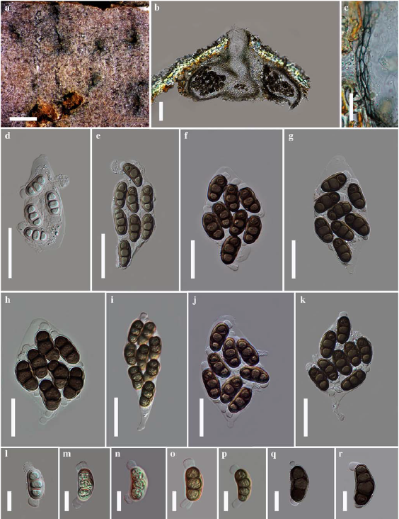
Fig. 2 Hapalocystis berkeleyi (MFLU 14-0798) a Ascomata on substrate b Cross section of ascomata c Peridium d–k Asci l–r Ascospores. Scale bars: a=1000μm, b=100μm, c=20μm, d-k=50μm, .l-r=20μm.
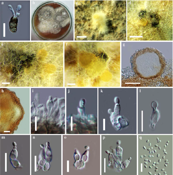
Fig. 3 “Phoma” like asexual morph of H. berkeleyi (MFLUCC 13- 0662) a Germinating ascospore b Culture on upper surface, growing on MEA c-f Conidiomata g Cross section of conidiomata h Peridium i–p Conidiophores, conidiogenous cells and conidia q Conidia. Scale bars: a=50μm, c=1000μm, d-f=500μm, g=100μm, h=20μm, i-p=10μm, q=5μm.