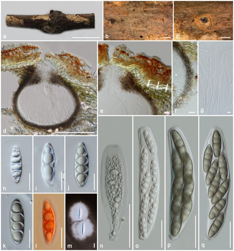
Massaria broussonetiae Samarak., Jian K. Liu & K.D. Hyde, sp. nov. Figure 1
MycoBank number: MB 843665; IndexFungorum number: IF 843665; Facesoffungi number: FoF 10807;
Etymology: The specific epithet refers to the host genus Broussonetia.
Holotype: HKAS 102402
Saprobic on a dead branch. Sexual morph: Ascomata 325–410 μm high × 285–340 μm diam. (M = 1.1 = 370 × 313 μm, n = 10), immersed in bark with erumpent neck, visible as black dots on the host surface, solitary, scattered or sometimes gregarious, compressed globose, coriaceous, brown to dark brown, with centrally opening ostiole. Ostiole centrally located, oblong, filled with periphyses. Peridium 27–35 μm (M = 31 μm, n =12) wide at the base and sides, 60–85 μm (M = 74 μm, n =8) wide around the ostiole, outer layer comprising reddish to dark brown, fused with host tissues, thin-walled cells of textura angularis, inner layer composed of hyaline, loosen, cells of textura angularis, green algae-like structures below the cambium. Hamathecium 1.8–2.7 μm (M = 2.2 μm, n = 15) wide, composed of numerous, dense, long, filamentous, branched, septate, trabeculate, round to blunt apex, pseudoparaphyses, embedded in a gelatinous matrix. Asci 115–150 × 33–53 μm (M = 192 × 42 μm, n = 25), 8-spored, bitunicate, fissitunicate, cylindrical or oblong, with a short pedicel, apically rounded. Ascospores 38–55 × 15–17.5 μm (M = 48.5 × 16.5 μm, n = 35), overlapping 2–3 seriate, fusoid to ellipsoid, hyaline, median eusepta, many guttules when immature, greenish brown, 3 septate when mature, slightly constricted at the medium septum, narrowly rounded to nearly acute at the ends, rough-walled, prominent, large rhomboid lumina in each cell, surrounded by a 26–88 μm (M = 48 μm, n = 10) wide, distinct mucilaginous sheath. Asexual morph: Undetermined.
Material examined: China, Guizhou, Guiyang, Guizhou Academy of Agricultural Sciences (GZAAS), on a dead branch of Broussonetia sp. (Moraceae) attached to the host, 22 July 2018, MC. Samarakoon, SAMC172 (HKAS 102402, holotype), isotype MFLU 19–2112. Additional sequences ITS XXXX, TUB2 XXXX.
Notes: Massaria broussonetiae morphology fits into the generic concept of Massaria as having globose, subglobose ascomata immersed in the substrate, centric ostiole, externally darkly pigmented peridium, indistinctly septate, branched, trabeculate pseudoparaphyses, bitunicate, fissitunicate asci, and large, ellipsoidal or fusoid, 3-septate ascospores with rhomboid or lenticular lumina in each cell and a distinct mucilaginous sheath. Interestingly, the bark surrounding the ascomata of our new collection is associated with green algae-like structures, which have not previously been described in any of the known Massaria species. The LSU sequence of M. broussonetiae is similar to that of M. parva WU 30553 (856/865; 99%), M. lantanae CBS 125592 (854/866; 99%), and M. ariae CBS 125589 (853/868; 98%), while RPB2 is similar to that of M. vomitoria WU 30607 (901/1046; 86%), M. platanoidea WU 30554 (901/1046; 86%), and M. inquinans WU 30527 (900/1046; 86%). The TEF1 sequence of M. broussonetiae is similar to that of M. pyri WU 30562 (702/750; 94%), M. aucupariae WU 30513 (699/750; 93%), and M. ariae WU 30510 (699/750; 93%). Combined gene phylogenies also show that M. broussonetiae clusters as a distinct clade, and here we introduce it as a new species.

Figure 1. Massaria broussonetiae (holotype HKAS 102402). (a) Substrate; (b,c) ascomata on substrate; (d) vertical section of ascoma; (e) ostiolar neck with green algae-like structures (in white arrows); (f) peridium; (g) paraphyses;.(h–m) ascospores (l in Congo Red, m in Indian ink); (n–q) asci. Scale bars: (a) = 1 cm; (b,c) = 500 µm; (d) = 200 µm; (n–q) = 50 µm; (e,f,h–m) = 20 µm, (g) = 5 µm.
