Marantokordyana oberwinkleriana M. Piepenbr., Maike Hartmann, T.A. Hofm. & M. Lutz, sp. nov.
MycoBank number: MB 832384; Index Fungorum number: IF 832384; Facesoffungi number: FoF. (Figs. 3a–c, 4, and 5)
Type on Goeppertia panamensis (Rowlee) Borchs. & S. Suárez (syn. Calathea panamensis Rowlee; Marantaceae). Panama, Chiriquí province, Dolega district, Los Algarrobos, trail to Río Majagua, 8° 29′ 22″ N, 82° 26′ 1″ W, 110 m asl., 10.8. 2018, leg. M. Piepenbring and M.U. Schmidt MP 5412 (holotype PMA 0123802, isotypes M 141363, UCH 11709).
GenBank: ITS = MN275897, LSU = MN275900.
Etymology: This species is named in honor of Franz Oberwinkler (1939–2018) who made important contributions to the knowledge of heterobasidiomycetes (comp. Piepenbring et al. 2019).
Description:
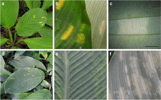
Leaf blades with scattered to numerous spots due to infection. Infected areas elongated rectangular and rounded at the ends, laterally delimited by veins of the leaf blade, not swollen, in surface view mostly (3–)4–10(−11) × (1.5–)2– 2.5(−3) mm (n = 10), sometimes larger by fusion, adaxially yellow to slightly orange colored, abaxially white surrounded by a ring of tissue of yellowish color, with white balls of basidia evident with a hand lens or a stereomicroscope. All the substomatal chambers of an infected area filled by fungal hyphae in dense stromata, hyphae protruding through the stomatal openings, developing balls of basidia mixed with paraphyses, one ball on the top of each stoma, balls of basidia easily breaking off and being dispersed.
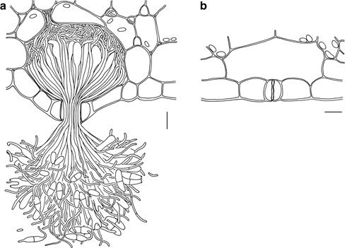
Substomatal chambers filled with fungal stroma formed by dense fungal hyphae, more voluminous than substomatal chambers without fungal stroma (Fig. 4), cellular details of fungal stroma difficult to distinguish, in very thin sections three types of fungal hyphae distinguishable, very thin (approx. 1 μm wide) hyphae close to host cells, thin (approx. 1.5 μm wide) hyphae mixed with the thick hyphae, and thick (approx. 3 μm wide) hyphae with swellings of up to 5 μm width at the base.
Balls of basidia spherical to globose, composed of basidia in different developmental stages and 1.5 μm wide paraphyses densely packed in the center and loosely exposed at the surface of a ball, (40–)55–80(−90) μm diam. (n = 20), not pigmented.
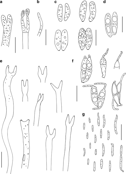
Basidia holobasidia, with basidial cell cylindrical, of variable length of up to at least 70 μm, sometimes shorter by retraction septa formed during sporulation, 3–4(–4.5) μm wide (n = 10), thinner after the liberation of the basidiospores, with two apical, straight or bent, elongated and often slightly swollen sterigmata of (2–)3.5–6.5(−7.5) × 1.5–2 μm (n = 20), with one basidiospore developing at the tip of each sterigma. After liberation of the basidiospores basidial cells empty, sometimes with scattered tiny remnants of cytoplasm, with scars evident at the tips of the sterigmata.
Basidiospores liberated singly or in pairs, at the moment of liberation one-celled but soon afterwards two-celled due to a central septum, cylindrical or slightly allantoid and slightly fusoid at the base, with a conspicuous hilum (scar) at the base of each basidiospore, (10–)11–16(−19) × 3–4.5(−6) μm (n = 30), hyaline, smooth, densely filled with oil drops or with homogeneous cytoplasm. The two basidiospores of one basidium conjugating by fusing or individual basidiospores germinating with about 1 μm wide hyphae originating at their tips or laterally and developing conidia on more or less evident sterigma-like outgrowths or cells of basidiospores directly developing conidia usually at their tips.
Conidia mostly one-celled, rarely with septum, rod-shaped to fusiform or slightly allantoid, with scar at the point of detachment, 3–8(−11) × (0.5–)1(−1.5) μm (n = 20), hyaline, smooth, germinating with thin hyphae or budding forming further conidia (yeast cells).
Further specimens examined: On Goeppertia panamensis. Panama, Chiriquí province, locality of the type, 12. 8. 2009, leg. M. Piepenbring and T.A. Hofmann M 409 (M 141366, UCH 11710). Same locality, 31. 8. 2009, leg. M. Piepenbring and T.A. Hofmann M 504 (M 141365, UCH 11711). Almost the same locality, 8° 29′ 18″ N, 82° 26′ 2″ W, 107 m asl., 27. 7. 2012, leg. M. Piepenbring and A. Krohn MP 5127 (M 141364, PMA 0123801). Panama, Chiriquí province, Gualaca district, close to Gualaca, canjilones de Gualaca, 8° 33′ 0.53″ N, 82° 18′ 3.44″ W, 133 m asl., 29. 7. 2018, leg. M. Piepenbring, N. Avila, R. Duarte, and M.U. Schmidt MP 5402 (UCH 11712; GenBank: ITS = MN275896, LSU = MN275899).

Fig. 3 Species of Marantokordyana. a–c M. oberwinkleriana on Goeppertia panamensis (MP 5127). a Infected plant in the field. b The upper side and the lower side of leaf blades with leaf spots. c A leaf spot seen from the lower side of the blade by a stereomicroscope. Each white dot is a ball of basidia. Scale bar = 2 mm. d–f M. boliviana on Goeppertia propinqua (CH 11). d Infected plant in the field. e The upper side of a leaf blade with leaf spots. f Leaf spots seen from the lower side of the blade. White dots are balls of basidia

Fig. 4 Marantokordyana oberwinkleriana on Goeppertia panamensis (MP 5412, holotype). a Transverse section of a leaf showing abaxial mesophyll, the abaxial epidermis, substomatal chamber filled with fungal cells (stroma), and a ball of paraphyses and basidia that liberated basidiospores single or in pairs. b Transverse section of a leaf showing abaxial mesophyll, the abaxial epidermis, and a substomatal chamber without fungal cells. Scale bars = 10 μm

Fig. 5 Marantokordyana oberwinkleriana (M 409, MP 5127). a Tips of young and sporulating basidia. b Tip of a paraphysis of a ball of basidia. c Basidiospores single or in pairs. d Two basidiospores connected by a conjugation bridge at their bases. e Tips of basidia after liberation of basidiospores. f Germinating basidiospores. g Conidia budding as yeasts. Scale bars = 10 μm
