Bleximothyrium ostiolatum Le Renard, Upchurch, Stockey & Berbee sp. nov.
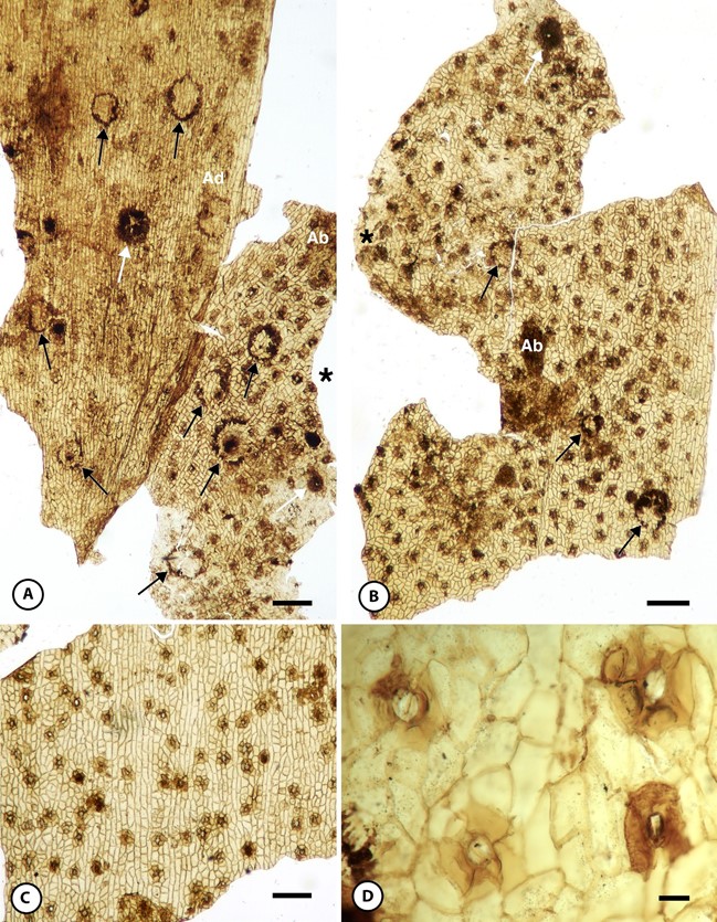
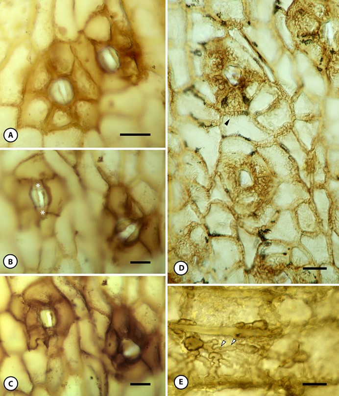
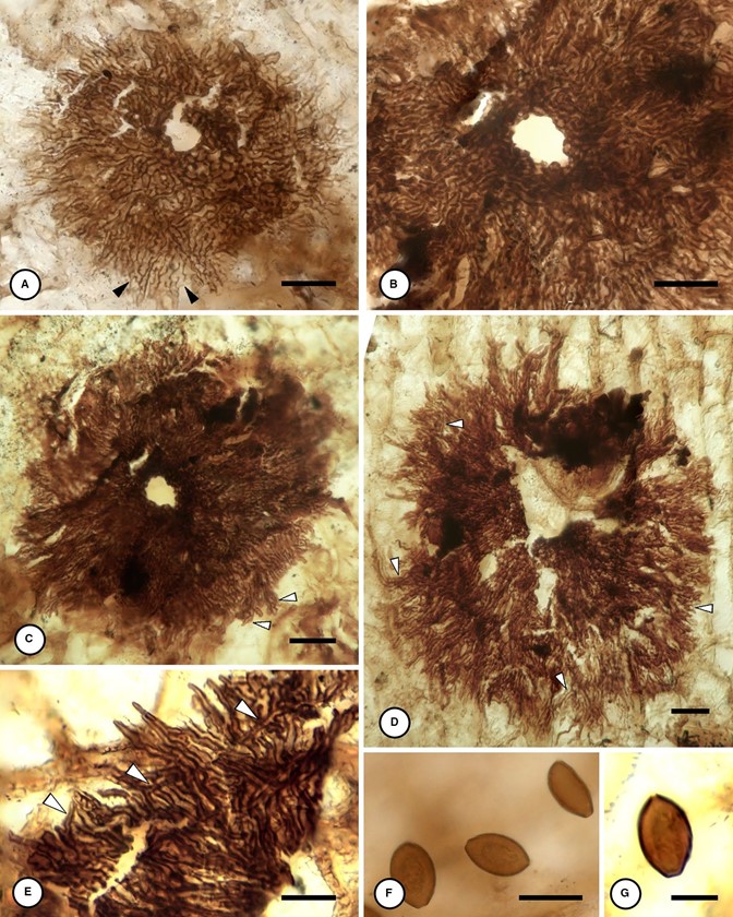
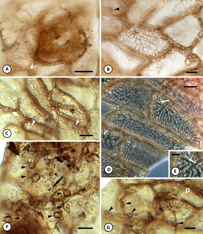
MycoBank number: MB 832578; Index Fungorum number: IF 832578; Facesoffungi number: FoF; Figs. 1A, 1B, 2D, 2E, 3A–3E, 4B, 4F, 4G
Specific diagnosis—Thyriothecium ostiolate, radiate, with isotomous branching, lower wall undifferentiated. Area near ostiole with pseudoparenchymatous hyphae. Surface hyphae connected to subcuticular mycelium via appressoria with pigmented penetration pegs. Subcuticular hyphae dendritic, forming extensive mycélium en palmettes (Ducomet, 1907).
Etymology—The specific epithet refers to the presence of an ostiole.
Holotype hic designatus—A specimen that consists of 16 sporocarps and a single, extensive mycelium on the surface of a single gymnosperm cuticle. Slide deposited at DMNS, under Accession 2019-55, slide #EPI 53111.
Locality—The Dutch Gap Canal (37.376°, −77.3593°; locality DMNH 128), James River, Virginia, USA.
Stratigraphic position and age—Potomac Group, lower Zone 1, Pseudofrenelopsis parceramosa bed; Aptian (Lower Cretaceous, 125–113 Ma).

FIGURE 1. Fossils of Bleximothyrium ostiolatum gen. et sp. nov. on Lower Cretaceous gymnosperm cuticle. (A, B) Overview of cuticle of single leaf colonized by B. ostiolatum. Two cuticle fragments attached at edges labeled with asterisks (*). White arrows indicate complete fungal scutella; black arrows indicate broken sporocarps; Holotype, DMNS EPI.53111. (A) Cuticle colonized by thyriothecia of B. ostiolatum; abaxial (Ab), adaxial (Ad) surface of same cuticle. Stomata are most numerous on abaxial side. Scale bar = 200 µm. (B) Smaller cuticle fragment showing only abaxial surface (Ab) in second piece of cuticle. Scale bar = 200 µm. (C, D) Gymnosperm cuticle similar to host of Bleximothyrium, not colonized by fungus, DMNS EPI.53112. (C) Overview of cuticle. Scale bar = 200 µm. (D) stomatal complexes. Scale bar = 20 µm.

FIGURE 2. Micromorphology of host plant cuticle and fungal colonization by Bleximothyrium ostiolatum gen. et sp. nov. (A) Two adjacent stomatal complexes from cuticle showing brown pigmentation of thick cuticle on subsidiary cells in absence of mycélium en palmettes. Three focal planes stacked; DMNS EPI.53112. Scale bar = 20 µm. (B, C) Uncolonized stomatal complexes. (B) Focal plane showing sunken guard cell cuticle; polar extensions labeled with asterisks (*); DMNS EPI.53112. Scale bar = 20 µm. (C) Focal plane showing four and five subsidiary cells; Holotype DMNS EPI.53111. Scale bar = 20 µm. (D) Mycelium of B. ostiolatum above epidermis and below cuticle; hyphae cover stomatal complexes and are in focus in troughs between adjacent pavement cells. Hyphae branch dichotomously before going out of focus, black arrowhead; Holotype DMNS EPI.53111. Scale bar = 20 µm. (E) Subcuticular mycelium showing irregular, lobed cells connected to surface mycelium by dark penetration pegs (white arrowheads). Holotype DMNS EPI.53111. Scale bar = 10 µm.

FIGURE 3. Morphology of scutella in Bleximothyrium ostiolatum gen. et sp. nov. Holotype DMNS EPI.53111. (A) Small mature sporocarp, showing radiate scutellum produced by hyphae branching with successive isotomous dichotomies (black arrowheads); ostiole is surrounded by pseudoparenchyma; eight focal planes (f.p.) stacked. Scale bar = 10 µm. (B–D) Mature large thyriothecia. (B) Central area of pseudoparenchyma near ostiole transitions to radiating hyphae in middle zone; eight f.p. stacked. Scale bar = 10 µm. (C) Overview of thyriothecium in (B). (C, D) Distal hyphae radiate, undulate, curve, and overlap, becoming tangled at margin (white arrowheads); from three f.p. stacked. Scale bars = 20 µm. (E) Distal margin of mature scutellum showing tangled distal hyphae (white arrowheads), with tapering hyphal tips. Scale bar = 10 µm. (F, G) Conidia of associated unknown fossil taxon showing blunt scar on one end and apical germ pore, each from three f.p. stacked. Scale bars = 10 µm in F; 5 µm in G.

FIGURE 4. Pattern of host colonization by Bleximothyrium ostiolatum gen. et sp. nov. (A–D) Subcuticular mycélium en palmettes follows contours of outer surface of host epidermal cells. Holotype DMNS EPI.53111. (A) Flat, densely branching palmettes proliferate under cuticle, over stomatal com- plex and across outer surface of epidermal pavement cell walls (white arrowhead). Scale bar = 20 µm. (B, C) Palmettes appressed against outer surface of epidermal cells and (B) in grooves between adjacent host cells (indicated with white asterisks *). Appressorium on cuticle surface (black arrowhead). Scale bar = 10 µm. (C) Flattened palmettes coating underside of cuticle (white arrowheads). Scale bar = 20 µm. (D) Extensive network of palmettes bearing occasional penetration pegs (white arrow). Phase contrast; scale bar = 10 µm. (E) Inset from (D) showing palmettes with penetration peg (white arrow). Scale bar = 5 µm. (F, G) Superficial mycelium with swollen appressoria (black arrowheads), showing penetration peg as black or hyaline dot in center. (G) Appressoria and their penetration pegs can be traced through cuticle to mycélium en palmettes (p). Scale bars = 10 µm.
