Lophiostoma ravennicum Tibpromma, Camporesi & K.D. Hyde.
Index Fungorum number: IF550884, Facesoffungi number: FoF00389; Fig. 2
Etymology – Refers to the name of the province in Italy where the fungus was collected.
Holotype – MFLU 14–0692
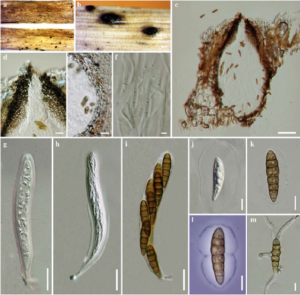
Saprobic on decaying grass stems of Juncus sp. Sexual morph Ascomata 211 – 282 μm high × 121 – 187 μm diam. (x̄ = 244 × 158 μm, n = 5), superficial, solitary, scattered, black, globose to subglobose, not easy to remove from host, neck long, black. Peridium 10 – 27 μm, 1 – layered, composed of small, light brown to dark brown, thin-walled cells of textura angularis. Hamathecium comprising numerous, 1.1 – 1.9 μm wide, long cellular, septate, hyaline, pseudoparaphyses, branching above the asci. Asci 55 – 70 × 9 – 11 μm (x̄ = 64 × 10 μm, n = 10), 8 – spored, bitunicate, fissitunicate, narrowly cylindrical, short pedicellate or apedicellate, apically rounded, with an ocular chamber. Ascospores 18 – 21 × 4 – 6 μm (x̄ = 19 × 5 μm, n = 15), 1 – 2 – seriate, brown, ellipsoidal-fusiform, narrowly fusoid with rounded ends, or fusiform, usually 6 – septate, the cells above central septum often broader than the lower ones, with mucilaginous sheath, smooth-walled. Asexual morph Undetermined.
Material examined – ITALY, Ravenna Province, Marina Romea, on stems of Juncus sp. (Juncaceae), 28 November 2013, E. Camporesi IT 1544 (MFLU 14–0692, holotype), ex-type living culture, MFLUCC 14–0005; GenBank ITS: KP698413; LSU: KP698414; SSU: KP698415.
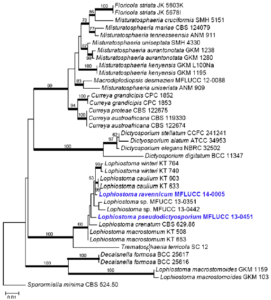
Notes – The molecular data confirms that our strain groups within the genus Lophiostoma (Fig. 1). Lophiostoma ravennicum however, differs in having ellipsoidal-fusiform, usually 6 – septate ascospores, with thick mucilaginous sheath and forms an individual lineage in the phylogenetic tree (Fig. 1).

Fig. 1 Phylogram generated from Maximum likelihood (RAxML) analysis based on LSU sequence data of Lophiostomataceae. Maximum likelihood bootstrap support values greater than 50 % are indicated above or below the nodes, and branches with Bayesian posterior probabilities greater than 0.95 are given in bold. The ex-types (reference strains) are in bold; the new isolates are in blue. The tree is rooted with Sporormiella minima CBS 524.50.

Fig. 2 Lophiostoma ravennicum (holotype) a, b Ascomata c Cross section of ascoma d Ostiole e Peridium f Pseudoparaphyses g–i Asci. j–l Ascospores m Germinating ascospore. Scale bars: a = 500 μm, b = 100 μm, c = 50 μm, d – e = 10 μm, f = 2 μm, g – i = 10 μm, j – m = 5 μm.
