Lophiostoma pseudodictyosporium Qing Tian, Camporesi & K.D. Hyde.
Index Fungorum number: IF550887, Facesoffungi number: FoF: 00403; Fig. 1
Etymology – In reference to the dictyosporous conidia similarity of the taxon to Dictyosporium.
Holotypus – MFLU 14–0737
Saprobic under peridermor semi-immersed in woody plant substrates Colonies growing on dead branch of Spartium junceum. Conidiomata pycnidial, on the upper surface of stem, (130–) 155 – 180 μm high × (145–) 160 – 195 μm diam. (x̄ = 165 × 180 μm, n = 10), solitary, scattered, superficial, globose to subglobose, black. Peridium thin at the apex, base and sides 12 – 15 μm (x̄ = 13.5 μm, n = 7), a single layer, composed of brown to black thin-walled cells of textura angularis. Conidiogenous cells (2–) 3 – 4.5 μm broad (x̄ = 3.5 μm, n = 10), holoblastic, integrated, smooth, brown, producing a branched conidium at the tip, cup-shaped or doliiform. Conidia (8–) 10 – 15 μm diam. × 12 – 26 (–32) μm high (x̄ = 12.5 × 23.5 μm, n = 10), uniformly medium brown, complanate, dictyosporous conidia, regularly consisting of 6 – 8 rows of cells, each rows comprising 5 – 7 cells, 3 – 3.5 μm wide.
Culture characters – Conidia germinating on PDA within 24 h and germ tubes arising from both ends. Colonies growing slowly on PDA, reaching a diam. of 1 cm after 30 days at 16 °C, velvety, radiating towards the edge. Mycelium initially hyaline and light pink at the margin, after 30 days, dense, filamentous, reverse black, pigments produced.
Material examined – ITALY, Province of Forlì-Cesena [FC], Fiumicello, Premilcuore, on stem of Spartium junceum (Fabaceae), 6 March 2013, E. Camporesi (MFLU 14–0737, holotype); ex-type living culture, MFLUCC 13–0451. GenBank ITS: KR025858; LSU: KR025862.
Notes – Lophiostoma is a morphologically well-studied genus (Barr 1990; Chesters and Bell 1970; Holm and Holm 1988; Hyde et al. 2013; Mugambi and Huhndorf 2009; Yuan and Zhao 1994; Zhang et al. 2012a, b). Lophiostoma was introduced by Cesati and De Notaris (1863), while Tanaka and Harada (2003a) typified L. macrostomum which was characterized by immersed to erumpent ascomata, with a cylindrical or crest-like papilla and slit-like ostiole; this has been regarded as a prominent morphological character of Lophiostoma macrostomum (Chesters and Bell 1970; Holm and Holm 1988). Combined gene analysis of LSU and SSU sequence data indicated Lophiostoma to be polyphyletic, grouping into two monophyletic clades (Zhang et al. 2009a, b, c; Suetrong et al. 2009). The asexual morph of Lophiostoma has rarely been reported. In this study, we introduce an asexual morph taxon, Lophiostoma pseudodictyosporium, which is shown to be related to Lophiostoma based on analysis of LSU gene sequence data. Lophistoma dictyosporium is morphologically similar with the species of Dictyosporium, both having brown hand-like conidia. In Dictyosporium the conidia form directly on mycelia, while in Lophiostoma pseudodictyosporium the conidia form in pycnidia. In the phylogenetic analysis they clustered into different families, therefore , we place the new species, Lophiostoma pseudodictyosporium in Lophiostoma, Lophiostomataceae.
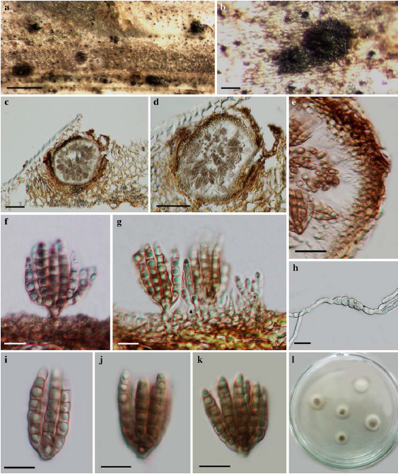
Fig. 1 Lophiostoma pseudodictyosporium (holotype) a Specimen material b Black conidiomata on the host c–e Vertical section of conidioma f–g Conidiogenous cells and developing conidia h Germinating ascospore i–k Conidia l Colony on PDA from above. Scale bars: a = 500 μm, b = 100 μm, c – d = 50 μm, e, h = 20 μm, f – g, i – k = 10 μm.